Melanoma is one of the most serious forms of skin cancer, known for its potential to spread quickly and become life-threatening if left untreated. However, the outlook for melanoma patients has changed dramatically over the past decade. With earlier detection, advanced therapies, and a deeper understanding of how melanoma behaves, survival rates have improved significantly.
This article explores how doctors determine melanoma prognosis, what influences survival outcomes, and what current statistics reveal about long-term life expectancy.
What Prognosis Means
A prognosis is an estimate of how a disease is likely to progress. In melanoma, it reflects a combination of medical findings — tumor characteristics, stage, patient health, and treatment response — to predict the chance of recovery or recurrence.
Prognosis doesn’t mean certainty; it provides a framework for understanding risk and planning treatment. Thanks to medical advances, many melanoma patients today live long, full lives after diagnosis, especially when the disease is caught early.
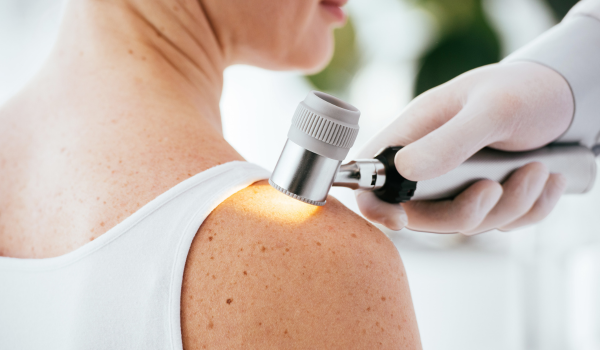
How Doctors Assess Prognosis
Doctors evaluate melanoma prognosis using a variety of clinical and laboratory factors. The most important is the stage of melanoma, determined after a biopsy and staging tests.
Other key considerations include:
-
Tumor thickness (Breslow depth): How deep the melanoma has grown into the skin.
-
Ulceration: Whether the skin surface over the tumor has broken down.
-
Mitotic rate: How quickly melanoma cells are dividing.
-
Lymph-node involvement: Whether cancer has spread to nearby lymph nodes.
-
Metastasis: Whether the cancer has spread to distant organs.
-
Patient factors: Age, sex, and overall immune health also affect outcomes.
All of these details help form a complete picture of prognosis and guide personalized treatment.
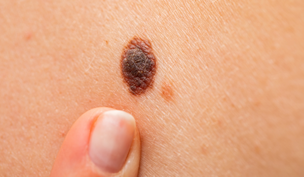
Tumor Thickness (Breslow Depth)
The Breslow depth is one of the strongest indicators of prognosis. It measures the melanoma’s vertical depth — from the top layer of the skin to the deepest point of cancer cells — in millimeters.
In general:
-
Less than 1 mm: Excellent prognosis; nearly 100% survival.
-
1–2 mm: High likelihood of cure with surgery.
-
2–4 mm: Increased risk of lymph-node involvement.
-
Greater than 4 mm: Higher risk of metastasis and lower survival rate.
Thinner tumors are usually detected earlier, which is why regular skin checks and prompt evaluation of suspicious moles are critical.
Ulceration and Mitotic Rate
Two microscopic features strongly influence melanoma outcomes:
-
Ulceration: When the surface skin over the melanoma breaks, it signals a more aggressive tumor. Ulcerated melanomas have a slightly lower survival rate compared to non-ulcerated ones of the same thickness.
-
Mitotic rate: This measures how many melanoma cells are dividing under a microscope. A higher rate suggests faster tumor growth and a greater chance of spreading.
These factors, combined with tumor depth, help doctors predict recurrence and tailor treatment intensity.
Lymph Node Involvement
The spread of melanoma to nearby lymph nodes marks a turning point in prognosis.
-
No node involvement (N0): Excellent outlook — most patients achieve long-term remission after surgery.
-
Microscopic involvement (N1a): Melanoma cells detected only by biopsy — moderate risk.
-
Macroscopic involvement (N1b, N2, N3): Nodes visibly enlarged or palpable — higher risk of recurrence or metastasis.
A sentinel lymph node biopsy (SLNB) is often performed to determine if microscopic spread has occurred, refining both staging and prognosis.
Metastasis and Distant Spread
When melanoma cells travel beyond regional lymph nodes to distant organs — such as the lungs, liver, brain, or bones — it is classified as Stage IV melanoma.
This stage historically carried a poor prognosis. However, with modern immunotherapies and targeted drugs, many patients now achieve durable remission or long-term survival. Some individuals live for 10 years or more with advanced melanoma, especially when treatment begins early after metastasis detection.
Melanoma Staging and Prognosis
Melanoma prognosis is closely tied to its stage at diagnosis:
| Stage | Description | 5-Year Survival Rate (Approx.) |
| 0 (In situ) | Confined to epidermis | ~100% |
| I | Thin, localized tumor | 95–99% |
| II | Thicker tumor, may be ulcerated | 80–90% |
| III | Lymph-node involvement | 50–80% |
| IV | Distant metastasis | 30–40% (improving with modern therapy) |
These statistics highlight why early detection is so vital — the difference between a curable Stage I melanoma and advanced Stage IV disease can be life-changing.
Prognostic Factors Beyond Staging
Beyond the official stage, several other biological and personal variables influence survival.
1. Age:
Older patients often have thicker or more advanced melanomas due to delayed detection. However, younger patients may develop more aggressive subtypes, such as nodular melanoma.
2. Gender:
Women generally have a slightly better prognosis than men, possibly due to differences in immune function or tumor location (women’s melanomas often occur on legs, men’s on the back).
3. Tumor Location:
Melanomas on the trunk or head have a higher risk of spreading than those on the limbs.
4. Immune System Health:
A strong immune response correlates with better outcomes. Immunocompromised individuals — such as transplant recipients — have reduced survival rates.
Molecular and Genetic Markers
Modern oncology uses genetic profiling to refine melanoma prognosis. Certain gene mutations affect how aggressively melanoma behaves and how it responds to treatment.
-
BRAF mutations: Present in about half of melanomas. These tumors respond well to BRAF/MEK inhibitor therapy.
-
NRAS mutations: Associated with more aggressive disease.
-
KIT mutations: Common in acral and mucosal melanomas, often treated with targeted drugs.
Identifying these mutations not only guides treatment but also provides insight into survival potential.
Impact of Treatment Advances
Over the past decade, survival rates have improved dramatically thanks to new therapies:
-
Immunotherapy (Checkpoint inhibitors): Drugs such as nivolumab, pembrolizumab, and ipilimumab activate the immune system to attack melanoma cells.
-
Targeted therapy: BRAF and MEK inhibitors shrink tumors in patients with specific mutations.
-
Combination therapy: Using both immunotherapy and targeted therapy often leads to long-term remission.
-
Adjuvant therapy: After surgery, high-risk patients receive medication to reduce recurrence.
These breakthroughs have transformed melanoma from one of the deadliest cancers into a highly manageable condition for many patients.
Survival Rates Over Time
According to the American Cancer Society and recent research, survival rates have steadily risen due to better screening and treatment.
-
In the 1980s, the overall five-year survival rate for melanoma was about 82%.
-
Today, it exceeds 93% across all stages combined.
-
For localized melanoma, survival approaches 99% when detected before spreading.
Long-term survival is increasingly common even in advanced melanoma, with some patients achieving “functional cures” through ongoing immunotherapy.
Understanding Relative Survival Rates
A relative survival rate compares people with melanoma to people without cancer of the same age and background.
For instance, a 90% five-year relative survival means that individuals with melanoma are 90% as likely to live five years as those without cancer.
This measure accounts for non-cancer causes of death and gives a clearer picture of melanoma’s direct impact on lifespan.
Recurrence Risk and Long-Term Monitoring
Even after successful treatment, melanoma can return — sometimes years later. The risk of recurrence depends on the original stage and tumor biology.
Recurrence types include:
-
Local recurrence: Near the original site.
-
Regional recurrence: In nearby lymph nodes.
-
Distant recurrence: In distant organs (metastasis).
Regular follow-up visits, skin checks, and imaging studies help catch recurrence early, when it’s still treatable.
Typical monitoring schedule:
-
Every 3–6 months for the first two years.
-
Every 6–12 months for years 3–5.
-
Annually thereafter.
Quality of Life and Survivorship
Modern treatment success means many melanoma patients become long-term survivors. However, they may face challenges such as:
-
Fear of recurrence or new lesions.
-
Skin sensitivity and scarring from surgery.
-
Side effects from immunotherapy (fatigue, inflammation).
Supportive care — psychological counseling, nutrition, exercise, and mindfulness — greatly enhances recovery and long-term quality of life.
Prognosis for Specific Subtypes
Different melanoma subtypes have unique behaviors that affect prognosis:
-
Superficial spreading melanoma: Most common; high cure rate when detected early.
-
Nodular melanoma: Grows rapidly; often diagnosed at a later stage.
-
Acral lentiginous melanoma: Appears on palms, soles, or under nails; common in darker skin; often diagnosed late.
-
Lentigo maligna melanoma: Usually develops in older adults on sun-damaged skin; slow-growing with excellent prognosis.
-
Mucosal melanoma: Rare and aggressive; poorer survival due to delayed detection.
Understanding subtype differences helps tailor treatment and anticipate outcomes.
Lifestyle and Long-Term Prevention
Prognosis isn’t just about treatment — lifestyle plays a vital role in long-term outcomes.
Protective habits include:
-
Daily broad-spectrum sunscreen (SPF 30+) use.
-
Avoiding tanning beds completely.
-
Wearing UV-protective clothing outdoors.
-
Eating antioxidant-rich foods to support immune health.
-
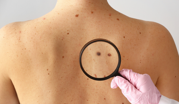
Performing monthly self-skin checks to catch new lesions early.
Prevention and vigilance can reduce recurrence risk and improve overall health for melanoma survivors.
Hope for the Future
Research continues to revolutionize melanoma care. Current studies focus on:
-
Personalized cancer vaccines that train the immune system to target melanoma cells.
-
Combination immunotherapies for resistant tumors.
-
Liquid biopsies that detect circulating melanoma DNA in blood before recurrence appears.
-
AI-driven skin monitoring tools that assist early diagnosis.
These innovations bring hope that melanoma survival will continue to climb, even in advanced stages.
Key Takeaways
-
Melanoma prognosis depends on tumor thickness, ulceration, lymph-node spread, and metastasis.
-
Early detection leads to survival rates near 100%.
-
Immunotherapy and targeted therapies have dramatically improved advanced-stage outcomes.
-
Regular monitoring and healthy habits help prevent recurrence.
-
Modern research offers more optimism than ever before.
Understanding your prognosis is not about fear — it’s about empowerment, awareness, and living proactively.
Final Thoughts
Melanoma survival rates have improved faster than nearly any other cancer, thanks to innovation and awareness. A diagnosis once considered devastating is now often treatable and manageable long term.
By learning the factors that affect prognosis and staying engaged in preventive care, patients can take control of their health journey. Early detection, timely treatment, and hope remain the strongest allies against melanoma.