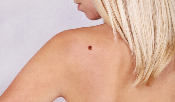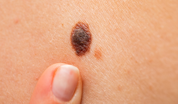
Melanoma is one of the most aggressive forms of skin cancer, developing in the pigment-producing cells known as melanocytes. These cells are responsible for giving color to your skin, hair, and eyes. When they begin to grow uncontrollably, melanoma can form and, if left untreated, spread rapidly to other parts of the body.
Understanding the types of melanoma is essential for early diagnosis and treatment. Each form behaves differently, looks different, and affects specific parts of the body. Here’s a detailed look at the main types—from the most common to the rarest.
Superficial Spreading Melanoma
Superficial spreading melanoma (SSM) is the most common type, accounting for around 70% of all melanoma cases. It usually begins as a flat or slightly raised patch on the skin that slowly expands outward before penetrating deeper layers.
This type often appears on the trunk in men and the legs in women, though it can develop anywhere. It’s characterized by irregular borders, multiple shades of brown or black, and a tendency to evolve gradually.
One key feature is the radial growth phase, where the tumor spreads horizontally within the top layer of the skin (the epidermis). During this stage, treatment outcomes are generally favorable if caught early.
Genetic studies often show BRAF or NRAS mutations in superficial spreading melanoma, which can influence treatment decisions involving targeted therapies.
Nodular Melanoma
Nodular melanoma (NM) is the second most common type and one of the most aggressive. Unlike other forms that grow outward first, NM grows vertically—deep into the skin layers—almost from the beginning.
It often appears as a raised, dome-shaped bump that can be black, blue, red, or even skin-colored. Because it grows quickly, it may ulcerate or bleed easily.
Nodular melanoma commonly appears on sun-exposed areas like the chest, back, or head. Its fast growth makes it harder to detect early, as it often bypasses the slower radial phase seen in superficial spreading melanoma.
Early detection is critical. Any new, rapidly growing mole or bump—especially one that looks different from the rest—should be evaluated by a dermatologist immediately.
Lentigo Maligna Melanoma
Lentigo maligna melanoma (LMM) develops from a precancerous condition called lentigo maligna, which can persist for years before turning invasive.
It’s most common in older adults, particularly those with a history of chronic sun exposure—like gardeners, sailors, or anyone who has spent much of their life outdoors.
You’ll typically find LMM on sun-damaged areas such as the face, ears, or neck. It appears as a flat, mottled patch with varying shades of brown, black, and sometimes gray.
The growth pattern is slow, often taking years before invading deeper skin layers. For this reason, early detection during routine skin checks offers an excellent prognosis.
LMM often carries KIT or NRAS mutations, which differ from the genetic signatures seen in other melanoma types.
Acral Lentiginous Melanoma
Acral lentiginous melanoma (ALM) is a rare but serious subtype that usually appears on the palms, soles of the feet, or under the nails (subungual melanoma).
It’s more common among people with darker skin tones, such as those of African, Asian, or Hispanic descent, though it can affect anyone.
Unlike other melanomas, ALM isn’t directly related to sun exposure. Instead, it often develops in areas with mechanical stress or trauma.
Clinically, ALM begins as a dark patch or streak under the nail or as an irregularly pigmented area on the palms or soles. Because it’s often mistaken for bruises or fungal infections, diagnosis is frequently delayed.
This delay makes ALM more likely to be diagnosed at an advanced stage, emphasizing the importance of awareness and early screening—especially for people of color.
Genetic analysis shows that ALM often lacks common BRAF mutations but may carry KIT mutations, influencing potential targeted treatments.
Desmoplastic Melanoma
Desmoplastic melanoma (DM) is one of the least common and most deceptive forms of melanoma. It tends to occur on sun-exposed areas, particularly the head and neck of older individuals.
This type is often non-pigmented (amelanotic) and may look like a scar or a firm skin-colored bump rather than a typical dark mole. Because of its subtle appearance, it’s often misdiagnosed as a benign lesion or even ignored.
Desmoplastic melanoma has a high tendency for nerve invasion (neurotropism), meaning it can spread along nerves, which complicates surgical removal. However, it’s less likely to spread to lymph nodes compared to other melanoma types.
Pathologists rely heavily on biopsy and immunohistochemical staining (such as S-100 protein) to confirm diagnosis.
Despite its challenges, DM often responds well to surgical excision with clear margins, and immunotherapy may play an increasing role in treatment.
Mucosal Melanoma
Mucosal melanoma (MM) originates in the mucous membranes that line body cavities such as the mouth, nose, throat, anus, or genital tract.
This form accounts for less than 2% of all melanomas but is extremely aggressive. Because mucosal tissues aren’t typically exposed to sunlight, the causes remain unclear, though genetic and environmental factors are likely involved.
Symptoms depend on the affected area. For example:
-
In the nasal cavity, it can cause nosebleeds or blockage.
-
In the mouth, it may present as a dark patch or ulcer.
-
In the genital area, it may cause bleeding or discomfort.
Mucosal melanoma is difficult to detect early due to its hidden location. As a result, it’s often diagnosed at an advanced stage, making treatment complex.
This melanoma type is frequently associated with KIT mutations, and clinical trials are exploring targeted therapies to improve outcomes.
Ocular (Uveal) Melanoma
Ocular melanoma develops in the uvea, the middle layer of the eye that includes the iris, ciliary body, and choroid. It’s the most common eye cancer in adults, though still rare overall.
Unlike skin melanoma, ocular melanoma is not linked to UV exposure in the same way. Instead, risk factors include light eye color, certain genetic syndromes, and older age.
Patients may experience blurred vision, flashing lights, or dark spots on the iris. Some cases are discovered incidentally during routine eye exams.
Treatment may involve radiation, laser therapy, or enucleation (removal of the eye) in severe cases.
Ocular melanoma behaves differently from cutaneous melanoma, with a unique tendency to spread to the liver rather than the lungs or lymph nodes.
Amelanotic Melanoma
Amelanotic melanoma (AM) lacks melanin pigment, which means it doesn’t appear dark or black like typical melanomas. Instead, it can look pink, red, or even skin-colored, making it extremely hard to recognize.
It may resemble eczema, a pimple, or a scar. Because of this, diagnosis is often delayed until the cancer has already advanced.
Amelanotic melanomas can occur in any part of the body and may arise from any of the other melanoma subtypes.
Dermoscopy and biopsy are vital tools for identifying these cases. Dermatologists often rely on the ABCDE rule (Asymmetry, Border, Color, Diameter, Evolving) and a high degree of suspicion to catch them early.
Rare and Variant Forms
Beyond the main subtypes, several rare or mixed forms exist. These include:
-
Spitzoid melanoma – resembles benign Spitz nevi but behaves malignantly.
-
Nevoid melanoma – mimics common moles yet carries invasive potential.
-
Animal-type (Pigmented epithelioid) melanoma – heavily pigmented and rare.
-
Polypoid melanoma – a nodular variant that grows outward like a stalk.
Each of these rare types presents diagnostic challenges. Pathologists use advanced molecular techniques to confirm their identity and guide treatment plans.
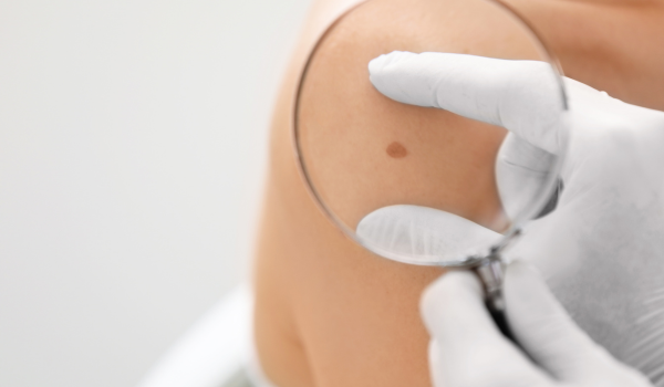
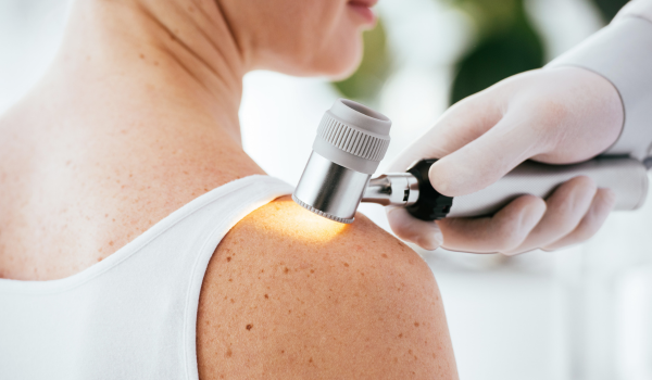
Diagnosis and Staging
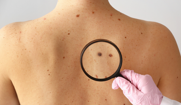
Diagnosing melanoma accurately requires a skin biopsy, where the suspicious lesion is removed and examined under a microscope.
Doctors evaluate several key features:
-
Tumor thickness (Breslow depth)
-
Ulceration or bleeding
-
Mitotic rate (how fast cells divide)
-
Lymph node involvement
-
Evidence of metastasis
These factors help assign a stage (0 to IV), guiding treatment and prognosis.
For deeper or advanced melanomas, imaging tests like PET-CT scans or MRIs may be used to check for spread to other organs.
Treatment Options
Treatment depends on the melanoma type, stage, and location. The most common approaches include:
-
Surgical removal: The first line of defense for localized tumors.
-
Immunotherapy: Drugs such as pembrolizumab and nivolumab help the immune system attack cancer cells.
-
Targeted therapy: Used when genetic mutations like BRAF, MEK, or KIT are detected.
-
Radiation therapy: Often applied for brain or bone metastases.
-
Clinical trials: Provide access to cutting-edge therapies and combination regimens.
Early detection remains the most powerful factor in improving survival rates, regardless of melanoma type.
Prevention and Early Detection
While not all melanomas are preventable, many can be avoided or caught early through proactive measures:
-
Protect your skin: Use broad-spectrum sunscreen daily, even on cloudy days.
-
Avoid tanning beds: Artificial UV light increases melanoma risk.
-
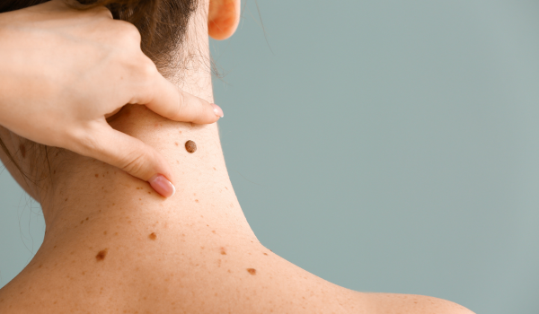
Perform self-checks: Examine your skin monthly for new or changing moles.
-
Schedule annual dermatology exams: Especially if you have a family history of melanoma or many moles.
Education and awareness are key. Recognizing the warning signs early can mean the difference between simple removal and life-threatening disease.
When to See a Doctor
Consult a dermatologist if you notice:
-
A new or changing mole
-
A spot that itches, bleeds, or doesn’t heal
-
Streaks under nails that don’t fade
-
Any skin changes that look different from your other spots
Prompt evaluation can save lives. Many melanomas are curable when detected at an early stage.
Key Takeaways
Melanoma is not a single disease but a group of cancers with diverse appearances and behaviors.
From superficial spreading melanoma—the most common—to acral lentiginous melanoma—a rare type that affects the palms and soles—each form requires unique diagnostic and treatment approaches.
The best defense is awareness, regular skin checks, and immediate medical attention for any suspicious change.