Melanoma is one of the most aggressive types of skin cancer, but not all melanomas behave the same. The stage of melanoma — determined by how deep the tumor has grown and whether it has spread — plays a crucial role in guiding treatment decisions and predicting outcomes.
Understanding these stages can help patients and families navigate diagnosis, treatment options, and what to expect next. Let’s break down each stage of melanoma and what it means for your care.
Why Staging Matters
Melanoma staging helps doctors assess how far the cancer has progressed. It determines whether the disease is confined to the skin or has reached lymph nodes and distant organs.
The American Joint Committee on Cancer (AJCC) developed the TNM system to describe melanoma stages:
-
T (Tumor): How thick and invasive the melanoma is.
-
N (Nodes): Whether the nearby lymph nodes contain cancer cells.
-
M (Metastasis): Whether the cancer has spread to other organs.
By combining these factors, doctors assign a stage from 0 to IV, guiding the best course of treatment and estimating prognosis.
Stage 0 – Melanoma In Situ
At this earliest stage, melanoma is confined to the outermost layer of the skin (epidermis) and has not invaded deeper tissues.
Characteristics:
-
Noninvasive; cancer cells remain in place.
-
No spread to lymph nodes or other organs.
-
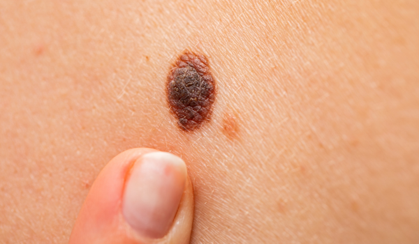
Often appears as an irregular mole or patch.
Treatment:
-
Surgical excision is usually curative. The lesion is removed with a small margin of normal tissue.
-
Follow-up involves regular skin checks to catch any new or recurring lesions.
Prognosis:
Nearly 100% survival rate when detected and treated early. The key is vigilance and routine skin monitoring.
Stage I – Early Invasive Melanoma
At Stage I, melanoma has penetrated below the epidermis into the dermis, but remains localized. It’s divided into sub-stages based on tumor thickness and ulceration.
Stage IA:
-
Tumor less than 1 mm thick.
-
No ulceration (open sore).
-
No lymph-node involvement.
Stage IB:
-
Tumor 1–2 mm thick without ulceration, or less than 1 mm with ulceration.
Treatment:
-
Wide local excision with margins of 1–2 cm of normal skin.
-
Sentinel lymph node biopsy (SLNB) may be recommended if the tumor is thicker than 0.8 mm.
Prognosis:
Five-year survival exceeds 95–99%. Regular follow-up is essential to detect any recurrence or new lesions.
Stage II – Regional Skin Spread
Stage II melanomas are thicker and may show signs of ulceration but still haven’t reached lymph nodes.
Sub-Stages:
-
IIA: 1–2 mm with ulceration, or 2–4 mm without ulceration.
-
IIB: 2–4 mm with ulceration, or over 4 mm without ulceration.
-
IIC: More than 4 mm thick with ulceration — indicating a higher risk of spread.
Treatment:
-
Surgical removal remains primary.
-
Sentinel lymph node biopsy is strongly recommended to check for hidden spread.
-
Adjuvant therapy (such as immunotherapy) may be considered if risk factors are present.
Prognosis:
Five-year survival rates range from 80–90%, depending on thickness and ulceration. At this stage, the focus is preventing local recurrence or early metastasis.
Stage III – Lymph Node Involvement
At Stage III, melanoma has spread to nearby lymph nodes or skin areas but not to distant organs.
Characteristics:
-
Cancer cells detected in one or more lymph nodes.
-
May spread to nearby skin or connective tissue (known as “in-transit metastases”).
-
Tumor thickness and ulceration can vary.
Treatment:
-
Surgery to remove the primary tumor and affected lymph nodes.
-
Adjuvant immunotherapy or targeted therapy (BRAF/MEK inhibitors if genetic mutations exist).
-
Radiation therapy may follow to reduce recurrence risk.
-
Clinical trials may be an option for advanced cases.
Prognosis:
Survival depends on how many lymph nodes are involved. On average, five-year survival rates are 50–80%. Ongoing research continues to improve treatment outcomes through advanced immunotherapies.
Stage IV – Metastatic Melanoma
This is the most advanced stage, where melanoma has spread beyond nearby lymph nodes to distant organs — commonly the lungs, liver, brain, or bones.
Symptoms depend on the affected organ:
-
Shortness of breath or chest pain (lungs).
-
Headaches, vision problems, or seizures (brain).
-
Fatigue or abdominal pain (liver).
Treatment:
Treatment aims to control cancer growth, extend life, and improve quality of life. Options include:
-
Immunotherapy: Checkpoint inhibitors (nivolumab, pembrolizumab, ipilimumab).
-
Targeted therapy: For patients with BRAF or MEK mutations.
-
Radiation therapy: To relieve symptoms or treat brain metastases.
-
Surgery: In selected cases to remove isolated metastases.
Prognosis:
Five-year survival rates have dramatically improved with modern treatments — now 30–40% for some patients. Early detection of spread and personalized therapies are key to extending survival.
The TNM Classification System
The TNM system provides a more detailed look at each stage:
-
T (Tumor Thickness):
-
T1: ≤1 mm
-
T2: 1.01–2 mm
-
T3: 2.01–4 mm
-
T4: >4 mm
Each “T” category also notes whether ulceration is present (a sign of more aggressive cancer).
-
-
N (Nodes):
-
N0: No lymph-node spread.
-
N1–N3: Increasing number and size of involved nodes.
-
-
M (Metastasis):
-
M0: No distant spread.
-
M1a–M1d: Spread to different distant sites (skin, lungs, liver, brain).
-
This system allows oncologists to personalize treatment and predict outcomes more precisely.
Prognostic Factors That Influence Outcome
Several factors beyond stage can influence melanoma outcomes:
-
Tumor thickness (Breslow depth): The deeper the tumor, the worse the prognosis.
-
Ulceration: Ulcerated melanomas indicate faster-growing, more aggressive disease.
-
Mitotic rate: The number of dividing cells under a microscope shows how quickly cancer grows.
-
Genetic mutations: BRAF, NRAS, or c-KIT mutations can impact treatment strategy.
-
Patient age and immune response: Younger, healthier patients often respond better to treatment.
Doctors use all these details alongside staging to develop a comprehensive management plan.
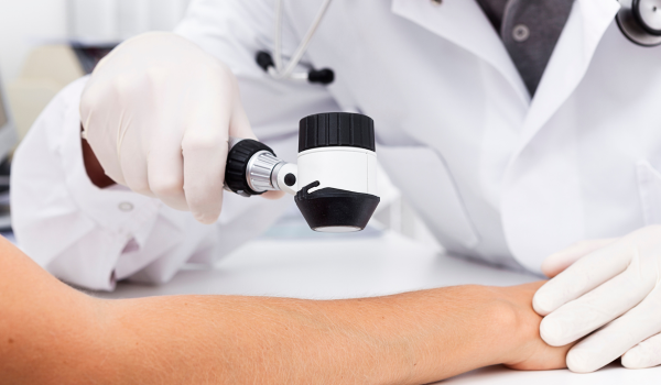
Sentinel Lymph Node Biopsy (SLNB)
This procedure is a cornerstone in melanoma staging. It identifies whether melanoma cells have spread to the nearest (sentinel) lymph node.
-
A radioactive dye is injected near the tumor site.
-
The first lymph node that absorbs the dye is surgically removed and analyzed.
-
If cancer cells are found, further lymph-node removal or adjuvant therapy may follow.
SLNB helps refine staging from Stage I–II to Stage III if spread is detected, improving treatment accuracy.
Treatment by Stage
Each melanoma stage has a unique treatment approach:
Stage 0:
-
Simple excision, no further therapy needed.
Stage I:
-
Wide local excision (1–2 cm margin).
-
Sentinel lymph node biopsy if >0.8 mm thick.
Stage II:
-
Surgery plus possible lymph node evaluation.
-
Immunotherapy or targeted therapy if high-risk features exist.
Stage III:
-
Tumor and lymph node removal.
-
Adjuvant immunotherapy or targeted therapy.
-
Possible radiation for high-risk areas.
Stage IV:
-
Combination of immunotherapy, targeted therapy, radiation, and surgery when appropriate.
-
Palliative care to manage symptoms and maintain quality of life.
Advances in Melanoma Treatment
Over the past decade, melanoma treatment has been revolutionized.
-
Immunotherapy: Checkpoint inhibitors like pembrolizumab and nivolumab enable immune cells to recognize and attack cancer.
-
Targeted therapy: For melanomas with BRAF mutations, drugs like dabrafenib and trametinib disrupt cancer growth pathways.
-
Combination therapy: Using both immunotherapy and targeted drugs has shown improved survival rates.
-
Oncolytic virus therapy: A new treatment using modified viruses to destroy cancer cells is being explored.
These therapies have transformed advanced melanoma from a deadly disease into one that is increasingly manageable.
Monitoring After Treatment
Even after successful treatment, patients must remain vigilant. Melanoma can recur years later.
Follow-up care includes:
-
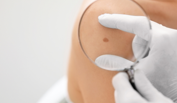
Dermatologic exams every 3–12 months, depending on stage.
-
Imaging (CT, PET, or MRI) if there’s risk of recurrence.
-
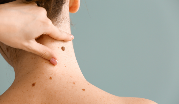
Patient self-checks for new moles or changes in old ones.
Patients should also adopt lifelong sun protection habits — sunscreen, protective clothing, and avoidance of tanning beds — to prevent recurrence and new cancers.
Living with Melanoma
Beyond physical health, melanoma affects emotional and mental well-being. Coping with a cancer diagnosis often involves anxiety, fear, or lifestyle adjustments.
Tips for living well with melanoma:
-
Join support groups or counseling for emotional support.
-
Maintain a healthy diet and regular exercise to strengthen immunity.
-
Discuss long-term side effects of treatment with your care team.
-
Stay informed about new clinical trials and evolving therapies.
A proactive mindset and medical follow-up can help patients lead fulfilling lives even after melanoma treatment.
Early Detection Saves Lives
The most powerful message about melanoma is that early detection changes everything.
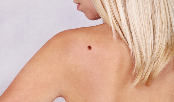
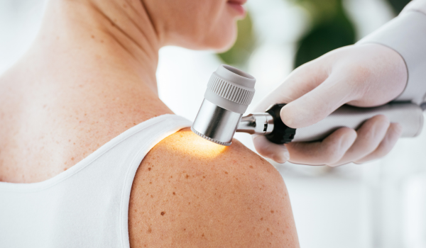
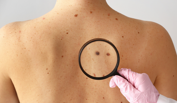
Checking your skin monthly using the ABCDE rule — Asymmetry, Border, Color, Diameter, and Evolution — can help identify melanoma when it’s most treatable.
If you spot a suspicious mole or spot that changes in shape, color, or texture, consult a dermatologist immediately. Early-stage melanomas are almost always curable, while advanced stages require more complex care.
Key Takeaways
-
Melanoma staging ranges from Stage 0 (in situ) to Stage IV (metastatic).
-
Staging depends on tumor thickness, lymph-node involvement, and metastasis.
-
Early stages are often curable with surgery; advanced stages need systemic therapy.
-
Immunotherapy and targeted treatments have dramatically improved survival.
-
Lifelong monitoring and prevention are crucial to reducing recurrence.
Understanding the stage empowers patients to take active roles in their treatment and recovery.
Final Thoughts
Melanoma staging is more than just a number — it’s the roadmap that guides every decision in your care journey. From localized to metastatic disease, knowing what each stage means helps patients, families, and clinicians make informed choices.
Early detection remains the strongest weapon against melanoma. Protect your skin, monitor your moles, and never ignore changes. When caught early, melanoma is one of the most preventable and treatable cancers of all.