Detecting melanoma early can save lives. Because melanoma is the most aggressive form of skin cancer, accurate and timely diagnosis is critical. Diagnosis typically begins with a clinical skin examination and proceeds through several specialized tests that help determine the type, depth, and spread of cancer.
Understanding these tests and procedures empowers patients to act quickly and confidently after noticing suspicious skin changes.
Recognizing Early Signs
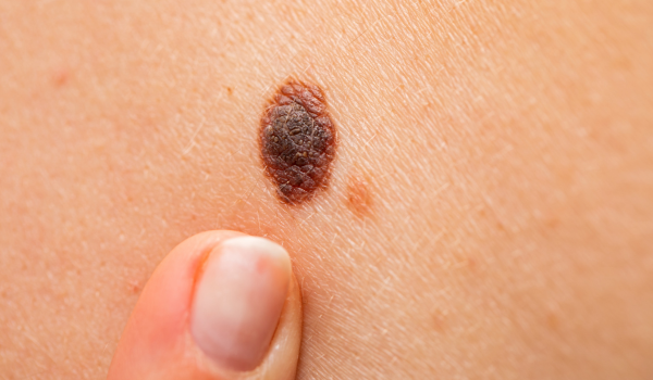
Diagnosis starts with awareness. Melanoma often begins as a mole that looks unusual or changes over time.
The ABCDE rule helps identify warning signs:
-
A – Asymmetry: One half of the mole doesn’t match the other.
-
B – Border: Edges are uneven, blurred, or jagged.
-
C – Color: Uneven shades of black, brown, red, or blue.
-
D – Diameter: Larger than 6 mm (about a pencil eraser).
-
E – Evolving: Changes in size, color, or texture over weeks or months.
Any mole that meets these criteria should be examined by a dermatologist as soon as possible.
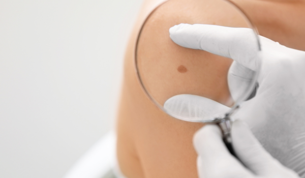
Initial Clinical Evaluation
A dermatologist begins with a comprehensive skin examination. Using bright light and magnification, they inspect the entire body — not just the visible mole — to look for other suspicious lesions.
During the evaluation, doctors also document:
-
Personal and family history of melanoma or other cancers.
-
Previous sun exposure or tanning-bed use.
-
Changes noticed by the patient.
The physician may photograph lesions for future comparison, especially when multiple or atypical moles are present.
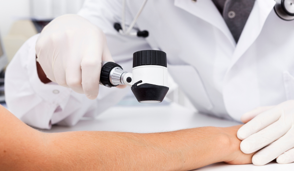
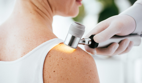
Dermatoscopy (Skin Surface Microscopy)
If a lesion appears suspicious, the next step is dermatoscopy, also called dermoscopy or epiluminescence microscopy.
A dermatoscope — a handheld device with polarized light and magnification — allows dermatologists to see subsurface skin structures invisible to the naked eye.
Benefits include:
-
Differentiating benign moles from malignant ones.
-
Detecting irregular pigment networks and vascular patterns.
-
Reducing unnecessary biopsies by improving diagnostic accuracy.
Dermatoscopy is painless, quick, and an essential part of modern skin-cancer detection.
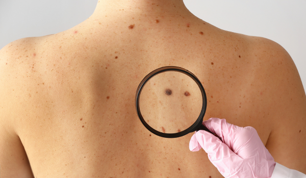
Digital Mole Mapping
For patients with numerous moles or a high risk of melanoma, doctors may recommend digital mole mapping.
This involves capturing high-resolution images of the entire body and specific close-ups of individual moles. Specialized software tracks these images over time to detect subtle changes.
Ideal for:
-
People with fair skin and many freckles.
-
Those with a strong family history of melanoma.
-
Patients who’ve previously had skin cancer.
By comparing old and new photos, dermatologists can detect melanoma at its earliest stage — before visible warning signs appear.
Skin Biopsy: The Definitive Test
A biopsy is the only way to confirm a melanoma diagnosis. It involves removing all or part of the suspicious lesion for microscopic examination.
There are several types of skin biopsies, chosen depending on the size and location of the lesion:
-
Excisional biopsy:
-
Removes the entire lesion plus a small margin of normal skin.
-
Preferred method for suspected melanoma.
-
-
Incisional (partial) biopsy:
-
Removes only part of a large lesion when full removal isn’t feasible.
-
-
Punch biopsy:
-
Uses a circular blade to remove a deeper skin core.
-
-
Shave biopsy:
-
Shaves off the top layers of the skin; used cautiously, as it may not capture tumor depth.
-
After removal, the sample is sent to a pathology laboratory for detailed analysis.
Histopathologic Examination
A pathologist — a doctor specialized in analyzing tissue — examines the biopsy under a microscope. This step determines whether melanoma cells are present and how advanced they are.
Key findings include:
-
Breslow thickness: Measures how deep the melanoma extends into the skin (in millimeters).
-
Clark level: Describes which layers of skin are affected.
-
Ulceration: Indicates whether the surface has broken down, a sign of aggressiveness.
-
Mitotic rate: Shows how quickly cancer cells are dividing.
These microscopic details are essential for accurate staging and prognosis.
Immunohistochemistry Testing
In some cases, the diagnosis is challenging — especially when melanoma resembles other pigmented skin conditions.
Immunohistochemistry (IHC) helps confirm the diagnosis by detecting specific proteins unique to melanoma cells, such as:
-
S-100 protein
-
HMB-45
-
Melan-A (MART-1)
IHC uses special antibodies and dyes that make melanoma cells stand out under the microscope, ensuring precision and minimizing misdiagnosis.
Molecular and Genetic Tests
Modern melanoma diagnosis goes beyond traditional microscopy. Genetic and molecular testing identifies mutations that can influence treatment.
Common mutations include:
-
BRAF (≈ 50 % of melanomas)
-
NRAS
-
KIT
Detecting these mutations helps doctors select targeted therapies, such as BRAF inhibitors (dabrafenib, vemurafenib) or MEK inhibitors (trametinib).
This approach personalizes treatment, offering better results and fewer side effects.
Sentinel Lymph Node Biopsy (SLNB)
Once melanoma is confirmed and deeper than 0.8 mm, doctors may perform a sentinel lymph node biopsy to check if cancer has spread.
Procedure:
-
A radioactive dye and blue dye are injected near the tumor.
-
The first lymph node (sentinel node) that absorbs the dye is surgically removed.
-
The node is examined under a microscope for cancer cells.
If the sentinel node contains melanoma, further lymph nodes may be removed or monitored closely. This procedure refines staging from local (Stage I–II) to regional (Stage III) disease.
Imaging and Scanning Tests
If there’s concern that melanoma has spread beyond the skin or lymph nodes, imaging tests help evaluate internal organs.
Common imaging studies include:
-
CT (Computed Tomography) scan: Detects spread to lungs, liver, or abdomen.
-
MRI (Magnetic Resonance Imaging): Especially useful for brain and spinal involvement.
-
PET (Positron Emission Tomography) scan: Identifies active cancer cells using a radioactive tracer.
-
Ultrasound: Evaluates lymph nodes near the tumor site.
These imaging methods help determine the extent of metastasis, confirming advanced-stage melanoma.
Blood Tests and Tumor Markers
Although no blood test alone can diagnose melanoma, doctors may order certain tests to support staging and monitor treatment response.
-
LDH (Lactate Dehydrogenase): Elevated levels can indicate advanced melanoma, especially when metastasis is present.
-
S-100 protein: Another marker that may rise in aggressive or metastatic cases.
-
Complete blood count (CBC) and liver function tests (LFTs) help assess overall health before treatment.
Regular blood monitoring is particularly important in Stage III–IV melanoma management.
Staging After Diagnosis
After all tests, doctors assign a stage (0–IV) based on tumor thickness, lymph-node involvement, and distant spread.
Staging determines:
-
Treatment approach (surgery vs systemic therapy).
-
Prognosis and survival rate.
-
Need for additional scans or adjuvant therapy.
Accurate staging transforms a biopsy result into a personalized action plan for each patient.
The Role of Pathology Reports
Every biopsy yields a detailed pathology report, which patients should review carefully with their dermatologist or oncologist.
The report includes:
-
Tumor type and subtype (e.g., superficial spreading, nodular, acral).
-
Breslow thickness, ulceration, mitotic index.
-
Margin status (whether the tumor was fully removed).
-
Recommendations for further testing or surgery.
Understanding the report helps patients stay informed and actively engaged in their care decisions.
Second Opinions and Expert Review
Because melanoma can mimic other skin conditions, obtaining a second pathology opinion from a specialist can be valuable — especially for ambiguous or rare subtypes.
Benefits of a second review:
-
Confirms or clarifies the diagnosis.
-
Ensures the most accurate staging.
-
Guides appropriate treatment choices.
Major cancer centers often provide expert consultations or review existing biopsy slides upon request.
Emerging Diagnostic Tools
Innovations continue to improve early detection and accuracy. Some promising developments include:
-
AI-based diagnostic imaging: Algorithms analyze mole photos to identify melanoma risk patterns.
-
Confocal microscopy: A non-invasive laser imaging method that visualizes skin cells in real time.
-
Liquid biopsy: Detects circulating tumor DNA (ctDNA) in the blood, offering early signs of recurrence.
These technologies aim to reduce unnecessary biopsies and catch melanoma before it becomes invasive.
Follow-Up After Diagnosis
Once melanoma is confirmed and treated, follow-up monitoring is crucial.
Typical schedule:
-
Every 3–6 months for the first two years.
-
Every 6–12 months for years 3–5.
-
Annually thereafter.
Each visit may include:
-
Skin examination for new lesions.
-
Lymph-node palpation.
-
Imaging or blood tests for higher-stage cases.
Consistent follow-up improves long-term survival and helps detect recurrences early.
Patient Awareness and Self-Checks
Patients play a key role in early detection and recurrence prevention. Regular self-checks allow you to recognize new or changing moles between appointments.
Tips for effective skin self-exams:
-
Use a full-length and hand mirror.
-
Check hard-to-see areas — back, scalp, soles, and under nails.
-
Track moles using smartphone apps or photos.
If you notice any new spot or mole that changes rapidly, seek medical advice promptly.
Emotional Impact of Diagnosis
Receiving a melanoma diagnosis can be emotionally overwhelming. Anxiety, uncertainty, and fear are common reactions.
Supportive care makes a difference:
-
Counseling or support groups can help manage stress.
-
Learning about treatment options restores control.
-
Sharing experiences with others encourages resilience.
Emotional well-being is a vital part of the healing journey and long-term recovery.
Prevention and Early Detection
Diagnosis and prevention go hand in hand. Many melanomas can be prevented by reducing UV exposure and maintaining skin health.
Protective strategies:
-
Apply broad-spectrum sunscreen (SPF 30+) daily.
-
Avoid tanning beds entirely.
-
Wear protective clothing and wide-brimmed hats.
-
Stay vigilant for suspicious skin changes year-round.
Early detection remains the most effective way to reduce melanoma deaths.
Key Takeaways
-
Melanoma diagnosis begins with skin examination and dermatoscopy.
-
Biopsy is the gold-standard test to confirm the disease.
-
Pathology, genetic, and imaging studies define the stage and guide treatment.
-
Regular follow-up and self-checks ensure early detection of recurrence.
-
Modern diagnostic tools — from AI to molecular testing — continue to improve survival outcomes.
Knowledge is power: understanding the diagnostic process helps patients make informed, confident decisions about their health.
Final Thoughts
Diagnosing melanoma involves a blend of visual assessment, microscopic analysis, and advanced imaging. Each step — from noticing a suspicious mole to genetic testing — contributes to a complete and accurate picture of the disease.
Early, precise diagnosis allows for prompt treatment and dramatically increases survival rates. Stay proactive: protect your skin, schedule annual dermatology visits, and never ignore changes that could signal melanoma.