Vitiligo is diagnosed through a combination of physical examination, blood testing, and skin biopsy. This condition results in the loss of skin pigment, appearing as lightened patches across various areas of the body. People experiencing symptoms such as pale spots on the skin or early graying of hair are often referred to dermatologists who specialize in skin disorders. These professionals conduct a series of evaluations to confirm vitiligo and rule out other possible conditions.
Vitiligo is classified as an autoimmune disease. This means the immune system mistakenly attacks the body’s own cells—in this case, melanocytes. These cells are responsible for producing melanin, the pigment that colors the skin. Both genetics and environmental factors are believed to influence the onset of vitiligo.
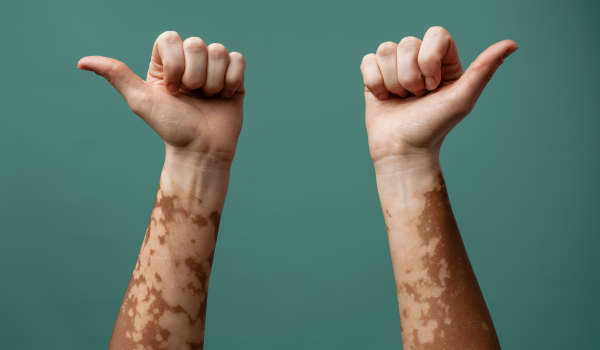
Self-check exam
While no home test can definitively diagnose vitiligo, individuals can monitor their own skin for early warning signs. Noticing pigment loss early can help speed up diagnosis and treatment planning.
common signs to look for include:
-
Lightened patches on the face, feet, or arms
-
Pale areas on the lips or around the mouth and nose
-
Premature whitening of scalp, beard, eyebrow, or eyelash hair
-
Itchy skin with visible discoloration
-
Eye color changes
In some cases, vitiligo may also increase sensitivity to sunlight or lead to hearing loss, as pigment-producing cells are found in the inner ear as well.
Identifying the pattern and location of depigmentation can help in determining the type of vitiligo:
-
mucosal vitiligo: affects the mucous membranes in the mouth, nose, or genital area
-
non-segmental vitiligo: the most common type; appears symmetrically on both sides of the body
-
segmental vitiligo: affects only one side or a specific area, typically in a localized form
Physical exam and medical history
A dermatologist will begin with a physical exam and collect detailed medical history. This includes questions about family history of vitiligo or other autoimmune diseases like diabetes.
important things to share with your provider:
-
Any recent skin rashes or unexplained discoloration
-
Home remedies, creams, or medications you’ve used
-
Known family history of skin pigment disorders
About one in five people with vitiligo has a close relative who also has the condition, making genetic factors relevant during diagnosis.
Blood tests
Blood tests are used to check for underlying autoimmune disorders that may coexist with vitiligo, especially thyroid disorders.
types of thyroid-related conditions:
-
hypothyroidism: marked by fatigue, weight gain, cold intolerance, and dry skin
-
hyperthyroidism: characterized by rapid heartbeat, weight loss, mood swings, and excessive sweating
Testing thyroid function helps assess levels of thyroid-stimulating hormone (TSH) to see if they are within a normal range.
Biopsy
A skin biopsy may be recommended when the diagnosis is uncertain. A dermatologist will remove a small sample of the affected skin for microscopic analysis.
key sign of vitiligo in a biopsy: absence of melanocytes, the pigment-producing cells
This procedure helps differentiate vitiligo from other skin conditions with similar symptoms.
Eye exam
Vitiligo can affect the eyelids and the hair in the eyebrow and lash areas. Though rare, it may also lead to complications in vision due to inflammation.
one possible eye condition linked to vitiligo is uveitis, which causes inflammation within the eye. An ophthalmologist may perform an exam to detect signs of this complication, especially if depigmentation is present on the face or around the eyes.
Hearing exam
Some individuals with vitiligo experience hearing changes due to immune system attacks on pigment cells in the ear. This type of hearing loss, called sensorineural hearing loss, stems from damage to the inner ear or auditory nerve.
audiologists or ENT specialists can perform hearing assessments if symptoms arise. Sensorineural hearing issues are among the more common, though still not universal, complications in vitiligo patients.
Screening for related conditions
Several skin conditions can cause white or lightened patches, so dermatologists use various tools to confirm that symptoms are caused by vitiligo and not another issue.
common conditions with similar symptoms include:
Tinea versicolor
This fungal infection leads to patches of discolored, flaky, or dry skin. Under a Wood’s lamp, the patches appear yellow, whereas vitiligo shows up as bright blue.
Albinism
Albinism is a genetic disorder affecting pigment in the skin, hair, and eyes. It’s usually diagnosed at birth. People with albinism have pale skin, white or very light blond hair, and often face vision issues like light sensitivity or misaligned eyes.
genetic testing may be advised if there is a family history of the condition.
Pityriasis alba
Mostly affecting children and teenagers, this condition causes pale, round, or oval patches. Under a Wood’s lamp, there’s no noticeable color change, distinguishing it from vitiligo.
Idiopathic guttate hypomelanosis
This condition presents as small white spots on sun-exposed skin, like the shoulders and arms. It's more common in older adults with fair skin. A biopsy can help confirm it by showing reduced melanin in the skin cells.
A quick review
Vitiligo is a chronic condition where the immune system attacks pigment cells, resulting in light patches on the skin or hair. Diagnosis typically involves a physical exam, medical history, blood tests, and sometimes a biopsy.
If symptoms extend to vision or hearing, further testing may be needed to identify related complications. Early diagnosis can help manage the condition and reduce discomfort or emotional distress caused by skin changes. Regular monitoring and communication with a healthcare provider are essential for long-term management.