Understanding Lymphoma Diagnosis
Lymphoma is a type of cancer that affects the lymphatic system, part of your immune defense network. Because its symptoms — like swollen lymph nodes, fatigue, fever, or weight loss — can mimic other common illnesses, accurate diagnosis requires multiple steps and specialized testing.
Doctors don’t rely on one single test to diagnose lymphoma. Instead, they combine physical exams, imaging scans, blood tests, biopsies, and laboratory analyses to determine whether cancer is present, what type it is, and how far it has spread.
Understanding these procedures can help patients feel more informed and less anxious when navigating their diagnostic journey.
Initial Evaluation
When lymphoma is suspected, the diagnostic process usually begins with a detailed medical history and physical examination.
Medical History
Your doctor will ask about:
-
Symptoms (when they started, how long they’ve lasted, and how severe they are).
-
Unintentional weight loss, night sweats, or prolonged fatigue.
-
Previous infections or autoimmune conditions.
-
Family history of lymphoma or other cancers.
-
Exposure to chemicals, radiation, or medications that might affect the immune system.
Physical Examination
The doctor checks for:
-
Swollen lymph nodes in the neck, armpits, or groin.
-
Enlarged spleen or liver (felt through the abdomen).
-
Skin changes, rashes, or visible lumps.
If the swelling has lasted more than two weeks and is painless, firm, or fixed, further testing is usually recommended.
Blood Tests
Blood tests cannot directly confirm lymphoma, but they provide important clues about your overall health and immune system function.
Common blood tests include:
-
Complete Blood Count (CBC): Measures red cells, white cells, and platelets. Abnormal results may suggest bone marrow involvement or anemia.
-
Erythrocyte Sedimentation Rate (ESR) or C-Reactive Protein (CRP): Detect inflammation, which can be elevated in lymphoma.
-
Liver and Kidney Function Tests: Assess whether the cancer has affected organ function.
-
Lactate Dehydrogenase (LDH): High levels may indicate tissue damage and aggressive lymphoma activity.
-
Viral Tests: Screening for viruses linked to lymphoma, such as Epstein-Barr virus (EBV), HIV, or Hepatitis B and C.
While blood tests alone don’t diagnose lymphoma, they help doctors evaluate severity, monitor progression, and plan safe treatment.
Imaging Scans
Imaging tests are vital for locating affected lymph nodes and organs and for determining how far the disease has spread. Each imaging method offers unique insights.
CT Scan (Computed Tomography)
A CT scan uses X-rays to produce detailed 3D images of your body. It helps identify:
-
Enlarged lymph nodes in the chest, abdomen, or pelvis.
-
Organ enlargement or tumor masses.
CT scans are often used both for initial staging and to monitor treatment progress.
PET Scan (Positron Emission Tomography)
A PET scan detects areas of high metabolic activity, which often indicates cancer. Patients are injected with a small amount of radioactive sugar (FDG). Lymphoma cells, being highly active, absorb more of it, lighting up on the scan.
PET scans are especially useful to:
-
Distinguish active disease from scar tissue.
-
Evaluate how well treatment is working.
-
Detect hidden cancer sites.
PET-CT Combination
In many hospitals, doctors use PET-CT scans, combining both technologies for the most accurate picture.
MRI (Magnetic Resonance Imaging)
MRI provides detailed images of soft tissues and is used when lymphoma may involve:
-
The brain, spinal cord, or bones.
-
Areas near vital organs, where precision matters most.
Ultrasound
Though less common for staging, ultrasound can detect superficial lymph nodes or organ enlargement, particularly in children or when radiation exposure needs to be minimized.
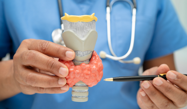
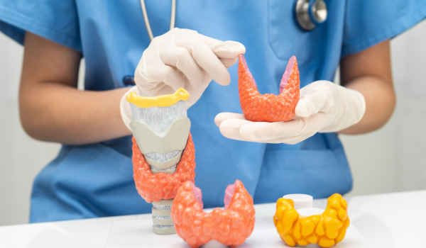
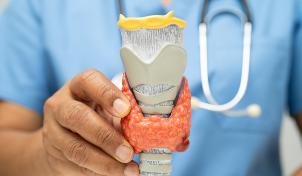
Lymph Node Biopsy
A biopsy is the cornerstone of lymphoma diagnosis. It’s the only way to confirm the presence of cancer cells and determine the exact type of lymphoma.
There are several biopsy techniques depending on the size and location of the lymph node.
Excisional Biopsy
-
Most accurate method.
-
Involves removing the entire lymph node under local or general anesthesia.
-
Allows pathologists to study cell structure, growth pattern, and genetic markers.
-
Considered the gold standard for diagnosis.
Incisional Biopsy
-
Removes only a portion of the lymph node when the full node can’t be accessed.
-
Often used for deep-seated nodes (e.g., in the chest or abdomen).
Core Needle Biopsy
-
A hollow needle extracts a small tissue cylinder from the node.
-
Minimally invasive and often guided by imaging (CT or ultrasound).
-
Suitable when surgery isn’t possible, though it may yield less tissue for analysis.
Fine Needle Aspiration (FNA)
-
Uses a thin needle to collect fluid or cells.
-
Helpful for preliminary evaluation but not sufficient for final diagnosis, as it doesn’t preserve tissue structure.
Once collected, the biopsy sample is sent to a pathology laboratory for detailed examination.
Pathology and Microscopic Examination
Pathologists examine the biopsy under a microscope to determine whether lymphoma is present and, if so, which type.
They look for:
-
Abnormal lymphocytes (like Reed-Sternberg cells in Hodgkin lymphoma).
-
Patterns of cell growth — clustered, diffuse, or nodular.
-
Presence of fibrosis, necrosis, or inflammation.
This microscopic evaluation distinguishes between Hodgkin lymphoma, Non-Hodgkin lymphoma, and other conditions that mimic them (such as infections or immune disorders).
Immunohistochemistry (IHC)
To confirm the diagnosis, pathologists use a technique called immunohistochemistry (IHC).
In IHC, special dyes and antibodies are applied to the tissue sample to detect specific proteins on the cell surface. These proteins act like identity markers that reveal what kind of lymphocyte the cancer originated from.
Common markers include:
-
CD20 – found on B cells.
-
CD3 – found on T cells.
-
CD30 and CD15 – typical of Hodgkin lymphoma.
By analyzing these markers, doctors can determine not only the lymphoma type but also how aggressive or indolent it might be.
Flow Cytometry
Flow cytometry is another test used to analyze cells from biopsy or blood samples. It measures how cells react to fluorescent antibodies targeting specific proteins.
This technique helps identify:
-
Whether cells are B-cell or T-cell origin.
-
Whether they are clonal (identical cancer cells) or normal.
-
How fast the cells are dividing.
Flow cytometry provides rapid, precise results and is essential for distinguishing lymphoma from other blood disorders.
Molecular and Genetic Testing
Advances in molecular biology allow doctors to study gene mutations, rearrangements, and chromosomal abnormalities within lymphoma cells. These tests help predict disease behavior and guide targeted treatment.
Common Techniques
-
FISH (Fluorescence In Situ Hybridization): Detects chromosomal translocations, such as t(14;18) in follicular lymphoma or t(8;14) in Burkitt lymphoma.
-
PCR (Polymerase Chain Reaction): Identifies specific gene rearrangements in B or T cell receptors.
-
Next-Generation Sequencing (NGS): Provides a detailed map of mutations for personalized therapy.
These insights help classify lymphoma subtypes and determine whether targeted drugs or immunotherapies may be effective.
Bone Marrow Examination
Because lymphoma can spread to the bone marrow, doctors often perform a bone marrow biopsy to assess disease extent.
Procedure
-
Done under local anesthesia, typically from the hip bone.
-
A needle is used to extract both liquid marrow (aspirate) and solid tissue (core biopsy).
-
The samples are analyzed for lymphoma cells, infection, or other abnormalities.
Bone marrow involvement helps determine the stage of lymphoma and can influence treatment decisions, especially in Non-Hodgkin lymphoma.
Lumbar Puncture (Spinal Tap)
If lymphoma is suspected in the central nervous system (CNS) — for example, in cases of aggressive B-cell lymphoma — a lumbar puncture may be performed.
Doctors insert a thin needle into the lower back to collect cerebrospinal fluid (CSF). The fluid is examined for cancer cells or abnormal proteins.
This test helps diagnose or rule out CNS lymphoma and decide whether intrathecal chemotherapy (drug delivery into the spinal fluid) is needed.
Staging the Disease
Once lymphoma is confirmed, doctors determine how far it has spread using the Ann Arbor staging system. This helps guide treatment planning.
| Stage | Description |
| Stage I | One lymph node region or a single organ involved |
| Stage II | Two or more regions on the same side of the diaphragm |
| Stage III | Lymph nodes on both sides of the diaphragm |
| Stage IV | Spread to organs such as liver, bone marrow, or lungs |
Additional letters like A/B indicate whether systemic symptoms (fever, night sweats, weight loss) are present.
Doctors may also use the Lugano classification, an updated system that incorporates PET-CT results for more precise staging.
Additional Laboratory Tests
Some additional tests may be done to evaluate how the body is coping with the disease or treatment:
-
Beta-2 Microglobulin: A protein that reflects tumor burden.
-
Uric Acid Levels: High levels may indicate rapid cell breakdown.
-
Serology Tests: Screen for autoimmune activity or coexisting infections.
These results help anticipate treatment-related complications and guide supportive care.
Why Multiple Tests Are Needed
Because lymphoma is not a single disease but a family of over 60 related cancers, accurate diagnosis depends on combining data from multiple tests.
For instance:
-
Imaging may show where the cancer is.
-
Biopsy reveals what kind of lymphoma it is.
-
Genetic testing shows how it behaves.
Together, these results allow oncologists to design a personalized treatment plan — targeting the specific lymphoma type, stage, and aggressiveness.
Preparing for Diagnosis
If your doctor suspects lymphoma, here are some ways to prepare for the diagnostic process:
-
Bring your medical records and a list of medications.
-
Ask questions about what each test involves and what it means.
-
Arrange support, as some procedures (like biopsies) may require rest afterward.
-
Take notes during appointments or bring a companion to help remember details.
Understanding each step can reduce anxiety and empower you to take an active role in your healthcare.
Emotional Impact and Support
Receiving a lymphoma diagnosis can be emotionally overwhelming. Uncertainty about test results, staging, or treatment can trigger stress and fear.
It’s important to remember that modern medicine offers highly effective treatments and excellent survival rates, especially for early-stage disease.
Consider:
-
Talking with a counselor or therapist.
-
Joining a lymphoma support group.
-
Connecting with patient advocacy organizations for guidance and community.
Emotional well-being is as vital as physical health during the diagnostic and treatment process.
Final Thoughts
Diagnosing lymphoma is a complex but precise process that combines advanced science and clinical expertise. From initial physical exams to genetic testing, every step helps doctors uncover the exact nature of the disease.
The goal isn’t just to find cancer — it’s to identify the specific type, stage, and biological profile, ensuring the most effective and personalized treatment plan.
By understanding these tests and procedures, patients can face the diagnostic process with confidence, clarity, and hope for recovery.