.png)
Why Diagnosis Matters
Cervical cancer can often be prevented or treated successfully if detected early. Most cases begin with precancerous cell changes in the cervix — changes that can be found through regular screening before they turn into invasive cancer.
Early diagnosis saves lives. It allows doctors to remove or treat abnormal tissue while it’s still localized, preventing spread to nearby organs.
Cervical cancer screening is not a single test but a combination of Pap smears, HPV testing, pelvic exams, and diagnostic follow-ups such as colposcopy or biopsy. Each step helps doctors move from simple suspicion to confirmed diagnosis.
Cervical Screening Overview
Screening aims to identify precancerous changes (CIN – Cervical Intraepithelial Neoplasia) caused by high-risk HPV infections before they progress. Screening does not diagnose cancer outright — instead, it flags abnormalities that require further testing.
Health authorities like the World Health Organization (WHO) and U.S. Preventive Services Task Force recommend:
-
Women aged 21–29 years: Pap test every 3 years.
-
Women aged 30–65 years: Pap + HPV co-testing every 5 years, or Pap alone every 3 years.
-
Women over 65 or post-hysterectomy may stop screening if previous results were normal.
These regular screenings are the most effective defense against advanced cervical cancer.
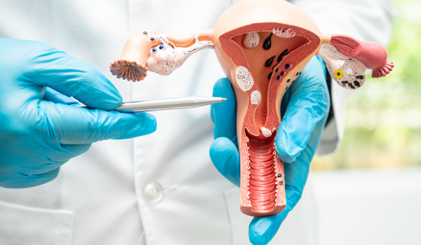
The Pelvic Exam
The diagnostic process usually starts with a pelvic examination. During this exam, the healthcare provider visually and manually assesses the vagina, cervix, uterus, and ovaries for abnormalities.
A speculum is inserted to open the vaginal canal, allowing inspection of the cervix. While not painful, slight pressure may be felt. Doctors look for visible lesions, inflammation, or unusual discharge.
If anything appears suspicious, they proceed with a Pap smear or other tests.
A pelvic exam alone cannot detect cancer cells — but it helps identify when additional screening is necessary.
Pap Smear (Pap Test)
The Pap test is the cornerstone of cervical cancer screening. It collects cells from the cervix to check for precancerous or cancerous changes under a microscope.
How it’s done:
-
A soft brush or spatula gently scrapes cells from the cervical surface.
-
The sample is preserved in liquid solution or placed on a glass slide.
-
It’s then sent to a laboratory for cytological analysis.
Results:
-
Normal: No abnormal cells found.
-
ASC-US / LSIL / HSIL: Indicate abnormal cell changes of varying severity.
-
Atypical cells suspicious for malignancy: Require further investigation.
If abnormal cells are detected, additional tests — such as HPV testing or colposcopy — will determine whether the changes are due to infection or early cancer.
HPV Test
Because almost all cervical cancers are caused by high-risk human papillomavirus (HPV), the HPV test is an equally critical tool. It detects viral DNA in cervical cells collected during a Pap or separate sample.
There are two main purposes:
-
Screening: For women aged 30 and older to identify high-risk strains before cell changes occur.
-
Triage: For women with abnormal Pap results to determine if HPV is responsible.
If the HPV test is positive for types 16 or 18 — the most dangerous strains — doctors may recommend immediate colposcopy even if Pap results are normal.
Co-Testing (Pap + HPV)
Co-testing combines both Pap and HPV tests for maximum accuracy. This approach improves detection rates of precancerous lesions while reducing unnecessary follow-ups.
A negative result on both tests means the risk of developing cervical cancer in the next five years is extremely low, which is why guidelines now allow a five-year interval between screenings for women 30–65.
Understanding Abnormal Results
Receiving an “abnormal” result does not mean cancer is present. It simply indicates that some cervical cells look different from normal under the microscope.
Common terms you might see:
-
ASC-US: Atypical Squamous Cells of Undetermined Significance.
-
LSIL: Low-grade Squamous Intraepithelial Lesion (mild changes).
-
HSIL: High-grade lesion (more serious, may progress).
-
AGC: Atypical Glandular Cells.
The next step depends on the grade. Low-grade changes often resolve naturally, while high-grade ones require closer evaluation through colposcopy or biopsy.
Colposcopy
When screening suggests abnormalities, doctors perform a colposcopy, a detailed visual examination of the cervix using a magnifying device called a colposcope.
Procedure:
-
The patient lies in a position similar to a Pap test.
-
The doctor applies a mild vinegar or iodine solution to highlight abnormal areas.
-
Suspicious regions turn white or change color.
-
Small tissue samples (biopsies) may be taken for laboratory analysis.
A colposcopy is typically painless, though mild cramping can occur. It helps determine whether the abnormal cells are precancerous (CIN 1–3) or invasive cancer.
Cervical Biopsy
The biopsy is the definitive diagnostic step. It involves removing a tiny piece of cervical tissue for microscopic examination. There are different types:
-
Punch biopsy: Removes small tissue samples from multiple sites.
-
Endocervical curettage (ECC): Scrapes cells from the cervical canal.
-
Cone biopsy (conization): Excises a cone-shaped section of tissue using a scalpel or loop (LEEP).
The biopsy helps pathologists classify cell abnormalities as:
-
CIN 1: Mild dysplasia, usually observed with follow-up.
-
CIN 2–3: Moderate to severe dysplasia — may require removal.
-
Carcinoma in situ or invasive carcinoma: Confirmed cancer.
A biopsy result is essential to confirm or rule out cervical cancer after abnormal screening.
LEEP and Cone Procedures
If a biopsy confirms precancerous lesions, doctors often use LEEP (Loop Electrosurgical Excision Procedure) or cold-knife conization to remove the affected tissue.
LEEP:
-
Uses an electrified wire loop to cut away abnormal areas.
-
Performed under local anesthesia.
-
Acts as both a treatment and a diagnostic sample.
Cold-knife conization:
-
Removes a deeper tissue cone in the operating room.
-
Used when invasive cancer is suspected or LEEP margins are unclear.
Both procedures can cure early-stage lesions and prevent cervical cancer from forming.
Imaging Tests
Once cervical cancer is confirmed, imaging tests determine how far it has spread — a process known as staging. Common imaging methods include:
-
Ultrasound: Evaluates the pelvis and nearby lymph nodes.
-
CT scan: Detects tumors in the abdomen, pelvis, or lungs.
-
MRI: Provides detailed views of soft tissue, helping assess tumor size and local invasion.
-
PET scan: Highlights active cancer cells using radioactive glucose.
These imaging studies guide treatment planning by showing whether the cancer is confined to the cervix or has metastasized.
Cystoscopy and Proctoscopy
In advanced cases, doctors may perform cystoscopy (to inspect the bladder) or proctoscopy (to check the rectum) to see if the cancer has spread beyond the cervix.
A thin scope with a camera is inserted to visualize the internal lining. These procedures are typically done under mild anesthesia and help with precise staging, especially before surgery or radiation.
Staging the Cancer
After diagnosis, doctors assign a stage (I–IV) to describe how far the cancer has spread. Staging is based on tumor size, lymph node involvement, and metastasis.
-
Stage 0: Carcinoma in situ (cells confined to the surface).
-
Stage I: Cancer limited to the cervix.
-
Stage II: Spread beyond the cervix but not to pelvic walls.
-
Stage III: Reached the pelvic wall or lower vagina.
-
Stage IV: Spread to bladder, rectum, or distant organs.
Staging determines treatment options such as surgery, radiation, or chemotherapy and helps predict prognosis.
Blood Tests
Although blood tests cannot diagnose cervical cancer directly, they support the process by evaluating overall health and organ function. Doctors may check:
-
Complete blood count (CBC): Detects anemia or infection.
-
Kidney and liver function tests: Ensure safety before chemotherapy or anesthesia.
-
Tumor markers (SCC antigen): Sometimes elevated in advanced squamous cell carcinoma, useful for monitoring treatment response.
HPV Genotyping
Some laboratories perform HPV genotyping, which identifies the exact HPV strain present in the cervix. Knowing whether the infection involves high-risk types 16 or 18 helps stratify patients by cancer risk and decide on follow-up intensity.
Women with persistent infection by these genotypes require closer surveillance, even when Pap results appear normal.
Self-Sampling for HPV Testing
Recent advances have made it possible for women to collect their own samples for HPV testing at home or in clinics. Self-sampling increases access for those who avoid pelvic exams due to cultural, emotional, or logistical barriers.
Studies show self-collected samples are nearly as accurate as clinician-collected ones, especially when analyzed with high-sensitivity molecular tests. Expanding these options could improve early detection rates worldwide.
When to See a Doctor
Women should not wait for screening schedules if symptoms appear. Warning signs that require medical evaluation include:
-
Abnormal vaginal bleeding (after sex, between periods, or after menopause)
-
Pelvic pain or pain during intercourse
-
Unusual vaginal discharge
-
Persistent back or leg pain in advanced stages
These symptoms don’t always indicate cancer but must never be ignored.
Emotional and Practical Support During Diagnosis
Being told you might have cervical cancer can trigger anxiety and fear. Understanding the diagnostic process helps reduce uncertainty.
Healthcare teams often include counselors or patient navigators who explain test results and next steps.
Support groups and online communities also provide encouragement. Remember: most abnormal results do not mean cancer, and even when it is present, early-stage disease is highly treatable.
Advances in Diagnostic Technology
Modern diagnostics are evolving rapidly. Artificial intelligence (AI)-assisted cytology, liquid-based cytology, and next-generation HPV assays are improving accuracy while reducing false positives.
Some systems use machine-learning algorithms to scan thousands of cervical cell images, identifying precancerous patterns faster than human eyes.
Emerging biomarkers and genetic profiling may soon help distinguish transient HPV infections from those likely to become cancerous — leading to more personalized care.
Global Efforts and Screening Accessibility
Despite medical advances, millions of women worldwide still lack access to basic cervical screening. Low-income countries account for over 85% of cervical cancer deaths due to limited healthcare infrastructure.
Global initiatives, such as the WHO Cervical Cancer Elimination Strategy, aim to:
-
Vaccinate 90% of girls against HPV by 2030.
-
Screen 70% of women at least twice in their lifetime.
-
Treat 90% of women with precancerous lesions.
Improving awareness, affordability, and access to diagnostic tools remains essential for reducing global mortality.
Key Takeaways
-
Early detection through Pap and HPV tests is the cornerstone of prevention.
-
Abnormal results require timely follow-up via colposcopy or biopsy.
-
Accurate staging through imaging guides treatment.
-
Self-sampling and AI tools are expanding access and accuracy worldwide.
-
Regular screening and HPV vaccination together could eliminate cervical cancer within decades.
.png)
.png)
.png)
.png)
.png)
.png)