Understanding Melanoma Types
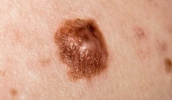
Melanoma is not a single disease but a group of skin cancers that develop from melanocytes — the pigment-producing cells responsible for skin color. While all types of melanoma start the same way, they behave differently depending on how deeply they invade, how quickly they spread, and where they occur.
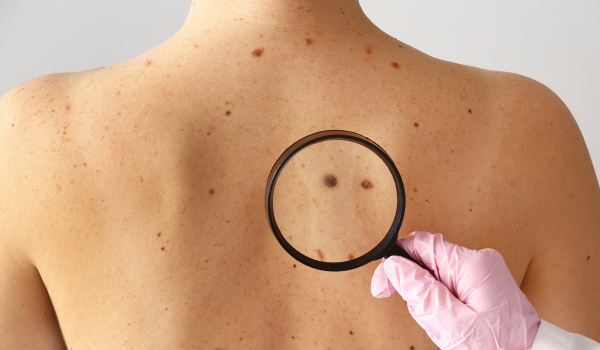
Recognizing the differences between types of melanoma is essential for early diagnosis and treatment. Some types are slow-growing and remain on the skin’s surface for months or years, while others are aggressive and can spread rapidly to other organs.
This article explores the main types of melanoma, their unique features, and what each means for diagnosis and treatment.
Superficial Spreading Melanoma
Superficial spreading melanoma (SSM) is the most common form, accounting for about 70% of all melanoma cases. It tends to develop on areas frequently exposed to the sun, such as the back in men and the legs in women.
Key characteristics:
-
Grows slowly across the top layer of skin before penetrating deeper.
-
Typically flat or slightly raised with uneven borders.
-
Shows mixed colors—brown, black, red, white, or blue.
-
Can arise from an existing mole or appear as a new spot.
Progression and risk:
SSM often develops from chronic sun exposure or repeated UV damage. Although it grows slowly, ignoring early signs allows it to invade deeper tissues and metastasize.
Treatment options:
-
Surgical excision with wide margins is usually curative when detected early.
-
Regular skin checks help detect new lesions or recurrences.
-
Sentinel lymph node biopsy may be considered for thicker tumors.
Nodular Melanoma
Nodular melanoma (NM) is the second most common type, accounting for about 15–20% of cases. It is also one of the most aggressive forms because it grows vertically, penetrating deep into the skin more quickly than other types.
Distinct features:
-
Appears as a raised bump or dome-shaped nodule.
-
Usually black or dark brown but may also be red, pink, or colorless.
-
Smooth, shiny surface that may ulcerate or bleed easily.
-
Commonly develops on the chest, back, or scalp.
Unlike superficial spreading melanoma, nodular melanoma often bypasses the slow radial growth phase, making early detection difficult. It can reach deeper layers within weeks or months.
Treatment:
-
Immediate surgical removal with wide margins.
-
Lymph node evaluation to check for spread.
-
Adjuvant therapies like immunotherapy if cancer has invaded lymphatic tissue.
Because nodular melanoma progresses rapidly, any suddenly growing, firm, or bleeding mole requires prompt medical evaluation.
Lentigo Maligna Melanoma
Lentigo maligna melanoma (LMM) primarily affects older adults and occurs in areas with years of sun exposure—most commonly the face, ears, and neck. It develops from a long-standing precancerous lesion called lentigo maligna (a type of melanoma in situ).
How it develops:
LMM begins as a flat, tan, or brown patch that gradually darkens and enlarges over several years. Its slow growth often delays diagnosis, as patients may mistake it for an age spot or sun damage.
Key signs:
-
Irregular shape with varying shades of brown or black.
-
Usually appears on sun-damaged skin of elderly individuals.
-
Flat or slightly elevated surface.
Treatment:
-
Surgical excision remains the gold standard.
-
Mohs micrographic surgery may be used to conserve tissue in delicate facial areas.
-
Topical immunotherapy (imiquimod) may be an option when surgery isn’t feasible.
Prognosis is excellent when detected early, but untreated LMM can evolve into invasive melanoma.
Acral Lentiginous Melanoma
Acral lentiginous melanoma (ALM) is a rare but serious form of melanoma that often goes unnoticed because it appears on parts of the body not typically exposed to sunlight—such as the palms, soles, and under fingernails or toenails.
Who is most affected:
ALM occurs in all skin types but is most common in people with darker skin tones (African, Asian, and Hispanic descent). It accounts for up to 50% of melanomas in people of color, even though overall melanoma rates are lower in these populations.
Typical features:
-
A dark streak under a nail (subungual melanoma).
-
Irregular patch on palms or soles that expands slowly.
-
May be mistaken for bruises, fungal infections, or calluses.
Treatment:
-
Surgical excision with adequate margins is essential.
-
Lymph node evaluation and imaging tests if invasion is suspected.
-
Targeted or immunotherapy in advanced cases.
Because acral lentiginous melanoma is often diagnosed late, awareness and early recognition are vital for improving outcomes.
Desmoplastic Melanoma
Desmoplastic melanoma (DM) is a rare subtype, making up less than 4% of melanomas, but it poses unique challenges in diagnosis.
It typically occurs on sun-exposed skin of older adults, particularly the head and neck, and often resembles a scar or firm lump rather than a pigmented mole.
Characteristics:
-
Usually skin-colored, pink, or gray instead of dark brown.
-
Hard, fibrous texture; may feel like a cyst or scar.
-
Grows deeply but slowly.
-
Often lacks pigment (amelanotic).
Treatment and outlook:
-
Surgical removal with wide margins is the main approach.
-
Radiation therapy may reduce local recurrence risk.
-
Immunotherapy may be added if metastasis occurs.
Because desmoplastic melanoma can mimic benign growths, biopsy of any persistent lesion is crucial for early detection.
Amelanotic Melanoma
Amelanotic melanoma (AM) is a subtype that lacks melanin pigment, meaning it appears pink, red, or skin-colored rather than the typical dark hue. This makes it easy to misdiagnose as a rash, wart, or inflamed lesion.
Why it’s dangerous:
Without pigmentation, amelanotic melanoma can go unnoticed until it reaches advanced stages. It may develop anywhere on the body and is often aggressive.
Common features:
-
Pink or reddish bump or patch.
-
May resemble eczema, scar tissue, or a pimple.
-
Rapidly changing size or thickness.
-
Occasionally painful or itchy.
Treatment:
-
Surgical removal with wide excision margins.
-
Lymph node biopsy to assess spread.
-
Systemic therapy (immunotherapy or targeted therapy) if advanced.
Awareness of non-pigmented melanoma signs is essential because “not all skin cancers are dark.”
Mucosal Melanoma
Mucosal melanoma forms on the mucous membranes—the moist linings of the body found inside the nose, mouth, throat, anus, or genitals. It’s extremely rare, accounting for only 1–2% of melanomas, but tends to be aggressive.
Symptoms vary depending on location:
-
Oral melanoma: dark patches or bleeding in the mouth.
-
Nasal melanoma: persistent congestion, nosebleeds, or sinus pain.
-
Genital melanoma: itching, pain, or color changes in the area.
Because mucosal surfaces are not exposed to UV light, researchers believe genetic mutations and immune factors play a stronger role than sunlight.
Treatment:
-
Surgical resection when feasible.
-
Radiation and immunotherapy to control local and systemic spread.
-
Close monitoring due to high recurrence risk.
Ocular (Uveal) Melanoma
Ocular melanoma develops in the eye’s uveal tract — the layer that includes the iris, ciliary body, and choroid. It is the most common eye cancer in adults, although it remains rare compared to skin melanoma.
Typical signs include:
-
Blurred or distorted vision.
-
Dark spot on the iris or inside the eye.
-
Flashes or floaters.
-
Change in pupil shape.
Treatment options:
-
Radiation therapy (plaque brachytherapy) for small tumors.
-
Surgical removal for large or advanced cases.
-
Immunotherapy or targeted drugs in metastatic stages.
Even though ocular melanoma is not caused by sun exposure, UV protection for the eyes (sunglasses with 100% UV filter) remains a wise preventive step.
Rare and Mixed Variants
There are several rare or mixed forms of melanoma that combine features of multiple subtypes or behave atypically:
-
Spitzoid melanoma: Resembles benign Spitz nevi, often pink or red, common in children and young adults.
-
Nevoid melanoma: Mimics a mole but is malignant underneath.
-
Polypoid melanoma: Appears as a protruding mass; grows rapidly.
-
Animal-type (equine) melanoma: Deeply pigmented and often slow-growing.
Because these types are less common, they often require pathological confirmation and specialized treatment plans from melanoma experts.
Amelanotic vs. Pigmented Types
While most melanomas produce melanin (dark pigment), amelanotic types lack color entirely. Both forms can be equally dangerous—what matters most is depth of invasion (Breslow thickness) and early detection.
Pigmented melanomas are easier to identify visually, whereas amelanotic melanomas demand careful clinical evaluation, often requiring dermoscopy or biopsy for confirmation.
Diagnosis Across All Types
Regardless of the subtype, early and accurate diagnosis is the cornerstone of melanoma treatment. Dermatologists use multiple techniques to identify the type and stage:
-
Dermoscopic examination to view skin structures beneath the surface.
-
Biopsy for microscopic analysis.
-
Breslow depth and Clark level measurements to assess tumor thickness and penetration.
-
Lymph node evaluation if spread is suspected.
-
Genetic testing to guide targeted therapies.
Once diagnosed, melanoma is staged (0–IV) to determine the most effective treatment strategy.
Treatment Overview by Type
Treatment depends on both the type and stage of melanoma but may include:
-
Surgery: Primary treatment for localized cases.
-
Immunotherapy: Drugs like pembrolizumab or nivolumab boost immune response.
-
Targeted therapy: Effective for BRAF or MEK mutations.
-
Radiation therapy: For head/neck lesions or palliative care.
-
Clinical trials: Testing novel vaccines, gene therapies, and combinations.
Prognosis is generally excellent for early-stage superficial types but worsens with late-stage nodular or mucosal melanomas.
Key Takeaways
-
Superficial spreading melanoma is the most common and easiest to treat when caught early.
-
Nodular melanoma grows quickly and demands urgent attention.
-
Acral and mucosal melanomas occur in less sun-exposed areas and are often diagnosed late.
-
Regular skin checks and awareness of unusual lesions are your best protection.
Early recognition of different melanoma types saves lives—knowledge truly is your best defense.
Final Thoughts
Melanoma may come in many forms, but they all share one message: early detection changes everything. Understanding the subtle differences among melanoma types helps patients, caregivers, and doctors respond faster and more effectively.
Whether it’s a slow-growing patch or a fast-developing lump, any skin change that looks or feels unusual deserves attention. In melanoma care, waiting is the most dangerous choice you can make.
Your skin is your first line of defense—watch it, know it, and protect it.