Understanding Melanoma Staging
Melanoma staging is a crucial process that helps doctors determine how far the cancer has spread, how aggressive it may be, and what treatment options offer the best outcomes.
Unlike other skin cancers, melanoma has a high potential to metastasize — or spread — beyond the skin to lymph nodes and internal organs.
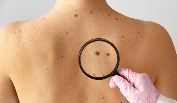
The stage of melanoma is identified after a biopsy and diagnostic tests such as sentinel lymph node mapping, imaging scans, or laboratory analyses.
Each stage corresponds to how deep the melanoma cells have penetrated and whether they have reached other parts of the body. Knowing your stage provides a clear roadmap for treatment and prognosis.
Why Staging Matters
Early-stage melanoma can often be removed with a simple surgical procedure. However, once cancer cells spread to the lymphatic system or distant organs, treatment becomes more complex and outcomes less predictable.
Staging helps oncologists:
-
Determine the extent of disease progression.
-
Choose appropriate therapies, from surgery to immunotherapy.
-
Predict chances of recurrence or remission.
-
Develop a personalized treatment plan and long-term monitoring schedule.
In short, staging is the foundation of every melanoma management decision.
How Doctors Determine the Stage
Doctors use several key factors to assign a melanoma stage, guided by the American Joint Committee on Cancer (AJCC) TNM system:
-
T (Tumor thickness and ulceration): Measured in millimeters using the Breslow depth scale.
-
N (Node involvement): Indicates whether cancer has spread to nearby lymph nodes.
-
M (Metastasis): Describes whether melanoma has reached distant organs like the lungs, liver, or brain.
Other considerations include mitotic rate (how quickly the cells divide) and ulceration (whether the tumor surface is broken). Combining these details gives doctors a clear clinical picture.
Stage 0 – Melanoma In Situ
Stage 0 is the earliest and least invasive form of melanoma. At this point, cancer cells are confined to the top layer of skin — the epidermis — and have not invaded deeper tissues.
Typical features:
-
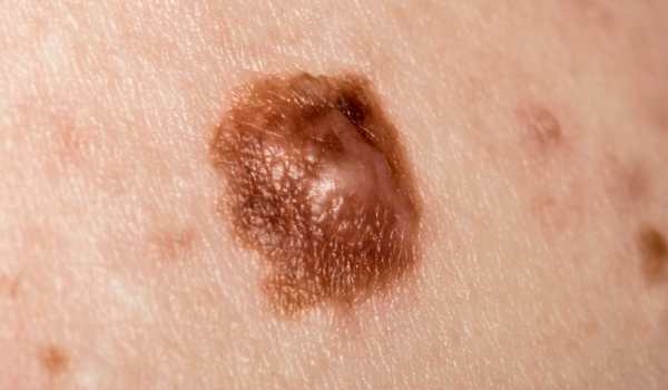
Appears as a flat or slightly raised mole.
-
Irregular borders or multiple shades of brown.
-
No spread to lymph nodes or other organs.
Treatment approach:
A dermatologist usually removes the lesion surgically with clear margins around it. No additional treatment is necessary, and the survival rate is virtually 100%.
Patients are encouraged to continue regular skin exams to detect any new changes early.
Stage I – Early Invasive Melanoma
Stage I melanoma indicates that the cancer has invaded deeper layers of the skin but hasn’t reached lymph nodes or distant sites.
Key traits:
-
Tumor thickness: ≤ 2 mm.
-
May or may not show ulceration.
-
No lymph node involvement.
Sub-stages:
-
IA: Tumor < 1 mm thick and not ulcerated.
-
IB: Tumor 1–2 mm or < 1 mm with ulceration.
Treatment options:
Surgical excision remains the gold standard. The surgeon removes the tumor and a margin of surrounding tissue (usually 1–2 cm).
In some cases, a sentinel lymph node biopsy may be performed to confirm that cancer hasn’t spread microscopically.
Prognosis:
Five-year survival exceeds 95% with proper removal and follow-up.
Stage II – Regional Skin Spread
Stage II melanoma is thicker and may show ulceration but still hasn’t reached the lymph nodes or distant organs.
Stage II A: 1–2 mm with ulceration or 2–4 mm without ulceration.
Stage II B: 2–4 mm with ulceration or > 4 mm without ulceration.
Stage II C: > 4 mm with ulceration (considered high-risk).
Treatment approach:
-
Wide surgical excision with broader margins.
-
Sentinel lymph node biopsy is typically recommended.
-
Adjuvant therapies may follow surgery to reduce recurrence risk.
Outlook:
Stage II has an excellent survival potential when detected early, though risk of recurrence is higher than in Stage I.
Stage III – Lymph Node Involvement
Stage III melanoma signifies that cancer has spread to nearby lymph nodes or adjacent skin but not yet to distant organs.
Common features:
-
One or more nearby lymph nodes test positive.
-
Satellite lesions or small tumor deposits may appear near the original site.
-
Tumor may or may not be ulcerated.
Sub-classification:
Stages IIIA through IIID depend on how many lymph nodes are involved and whether the cancer is ulcerated or microscopic.
Treatment approach:
-
Surgical removal of the primary tumor and affected lymph nodes.
-
Adjuvant immunotherapy (e.g., nivolumab, pembrolizumab).
-
Targeted therapy for BRAF mutation-positive tumors.
-
Radiation therapy for residual disease.
Prognosis:
Survival rates vary widely, ranging from 40 to 80%, depending on tumor thickness, ulceration, and the number of nodes involved.
Stage IV – Metastatic Melanoma
Stage IV is the most advanced form of melanoma. Here, cancer cells have traveled through the bloodstream or lymphatic system to distant organs such as the lungs, liver, brain, or bones.
Typical indicators:
-
Cancer found in distant skin, soft tissues, or internal organs.
-
Weight loss, fatigue, or neurological symptoms.
-
Elevated LDH (lactate dehydrogenase) levels in blood tests.
Treatment options:
-
Immunotherapy: Checkpoint inhibitors (e.g., nivolumab, pembrolizumab, ipilimumab) can stimulate the immune system to attack melanoma cells.
-
Targeted therapy: For BRAF and MEK mutations (vemurafenib, dabrafenib + trametinib).
-
Chemotherapy or radiation: Used less frequently but sometimes to relieve symptoms or control specific tumor sites.
-
Surgical removal of isolated metastases may be considered if feasible.
Prognosis:
Although historically poor, survival has improved dramatically thanks to modern immunotherapies. Many Stage IV patients now experience long-term remission.
The Meaning of Letters A, B, C, D
Each stage may include letters (A – D) that further define how extensive the disease is.
-
A: Thinner, non-ulcerated tumors or minimal lymph involvement.
-
B: Ulceration present or slightly thicker lesions.
-
C: Lymph node involvement with ulceration.
-
D: Widespread metastasis or multiple organ spread.
These letters provide more precise details, helping doctors fine-tune treatment.
Staging Systems Used Worldwide
There are two primary systems used to describe melanoma progression:
1. Ann Arbor System (originally for lymphoma but adapted for melanoma in some regions):
Divides stages based on lymph node and organ involvement.
2. Lugano Classification and AJCC TNM System:
The modern standard worldwide, incorporating tumor thickness, ulceration, lymph nodes, and metastasis for a complete picture.
Doctors also use Clark’s levels, which describe how deeply melanoma cells have penetrated the skin layers, but Breslow depth is now more predictive of prognosis.
Key Prognostic Factors
Besides staging, several elements influence how a person responds to treatment:
-
Tumor thickness (Breslow depth): The deeper it penetrates, the higher the risk.
-
Ulceration: Indicates a more aggressive tumor.
-
Mitotic rate: How fast the cancer cells divide.
-
Immune system health: Strong immunity often leads to better outcomes.
-
Location: Head, neck, or mucosal melanomas tend to have worse prognoses.
Understanding these details allows patients and doctors to take a proactive approach to treatment.
Treatment Options by Stage
Here’s how treatment typically progresses through the stages:
Stage 0–I:
-
Surgical excision only.
-
Regular dermatologic monitoring.
Stage II:
-
Wide excision ± sentinel lymph node biopsy.
-
Consider adjuvant immunotherapy.
Stage III:
-
Surgery + lymph node dissection.
-
Immunotherapy or targeted drugs.
-
Possible radiation.
Stage IV:
-
Systemic immunotherapy or targeted therapy.
-
Palliative radiation or surgery for symptom relief.
-
Clinical trials for novel therapies.
Innovative Treatments Changing Outcomes
Modern science is revolutionizing melanoma care.
-
Checkpoint inhibitors help the immune system recognize and destroy melanoma cells.
-
Targeted therapies block genetic mutations driving tumor growth.
-
Combination therapies offer stronger, longer-lasting responses.
-
Clinical trials test next-generation vaccines and gene-based treatments.
These advances have transformed melanoma from a near-fatal disease into one that can often be managed — or even cured — with early and precise intervention.
Monitoring After Treatment
Even after successful treatment, melanoma survivors require lifelong vigilance. Follow-up may include:
-
Dermatology visits every 3–6 months.
-
Periodic scans to detect recurrence.
-
Self-exams for new or changing moles.
-
Lifestyle adjustments such as sun protection and healthy diet.
Regular monitoring ensures that any recurrence is caught early, improving the chance of quick and effective management.
Living with Melanoma
Coping with melanoma involves physical and emotional challenges. Patients often face anxiety about recurrence and must balance long-term medical follow-up with daily life.
Support groups, therapy, and a strong social network can ease this journey. Many survivors find empowerment in sharing their stories, raising awareness, and adopting preventive habits that inspire others to take skin health seriously.
Key Takeaways
-
Melanoma staging defines how far cancer has spread and guides treatment.
-
Early stages (0–I) often require only surgery, while later stages may need immunotherapy or targeted therapy.
-
Regular follow-up is critical to prevent recurrence.
-
Advances in treatment have greatly improved survival and quality of life.
Knowledge of your melanoma stage gives you control — it helps you make informed choices and move forward with confidence.
Final Thoughts
Melanoma is serious but increasingly manageable with early detection and advanced treatments. Understanding what each stage means empowers you to act quickly, ask informed questions, and commit to follow-up care.
If you notice unusual skin changes or have already been diagnosed, remember: staging is not the end of your story — it’s the beginning of your path toward recovery.