.png)
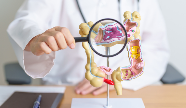
Introduction
Ulcerative colitis (UC) is a chronic inflammatory bowel disease (IBD) that affects the colon and rectum. Because its symptoms—such as diarrhea, abdominal pain, and rectal bleeding—can mimic other digestive conditions, a precise diagnosis is essential.
Doctors rely on a combination of patient history, physical examination, lab work, imaging, and endoscopic procedures to confirm UC and rule out similar diseases like Crohn’s disease, infections, or irritable bowel syndrome (IBS).
This article explores the tests and examinations doctors use to diagnose ulcerative colitis, why they matter, and what patients can expect during the process.
Medical History
The first step in diagnosing UC is a thorough medical history review. Doctors ask detailed questions about:
-
Symptoms: frequency of diarrhea, presence of blood or mucus in stool, abdominal pain patterns.
-
Duration: how long symptoms have persisted and whether they fluctuate.
-
Family history: relatives with IBD, autoimmune diseases, or colon cancer.
-
Lifestyle factors: diet, stress, medications, and smoking habits.
A strong family history or long-standing gastrointestinal issues may immediately raise suspicion of UC.
Physical Examination
After reviewing history, physicians perform a physical examination to detect visible or palpable signs of inflammation. This includes:
-
Checking for abdominal tenderness or bloating.
-
Evaluating for weight loss or malnutrition.
-
Assessing for anemia (pale skin, fatigue).
-
Looking for extraintestinal symptoms such as joint pain, eye inflammation, or skin rashes.
While the physical exam alone cannot confirm UC, it helps guide which diagnostic tests are most appropriate.
Blood Tests
Blood tests are crucial in identifying markers of inflammation and complications. Common tests include:
-
Complete blood count (CBC): detects anemia caused by chronic blood loss.
-
C-reactive protein (CRP): measures inflammation levels.
-
Erythrocyte sedimentation rate (ESR): another indicator of systemic inflammation.
-
Electrolyte levels: assess dehydration or imbalance from persistent diarrhea.
Blood work does not confirm UC on its own but helps doctors understand the severity of disease and rule out other conditions.
Stool Tests
Stool samples provide insight into infections and inflammation in the digestive tract. Doctors may order:
-
Fecal calprotectin test: measures inflammation proteins in stool, often elevated in UC.
-
Stool cultures: rule out bacterial, viral, or parasitic infections.
-
Occult blood test: checks for hidden blood in stool.
These tests are especially important in distinguishing UC from short-term infectious diarrhea.
Colonoscopy
The colonoscopy is the gold standard for diagnosing ulcerative colitis. During this test:
-
A long, flexible tube with a camera is inserted into the rectum.
-
The doctor examines the entire colon for redness, ulcers, or bleeding.
-
Biopsies (small tissue samples) are taken for microscopic analysis.
Key findings in UC include continuous inflammation starting from the rectum—a feature that distinguishes it from Crohn’s disease, which typically shows patchy involvement.
Flexible Sigmoidoscopy
Sometimes doctors use a flexible sigmoidoscopy, which examines only the rectum and lower part of the colon.
-
This test is less invasive than a full colonoscopy.
-
It is often performed when the colon is too inflamed for a complete examination.
-
Like colonoscopy, it allows for biopsies.
Though it provides a limited view, sigmoidoscopy is valuable for diagnosing UC in urgent or severe cases.
Imaging Studies
Imaging helps doctors assess complications or rule out other diseases. Common imaging tests include:
-
CT scan: detects abscesses, perforations, or severe inflammation.
-
MRI scan: especially useful for evaluating soft tissues and detecting complications.
-
X-rays: may be used in emergencies to check for toxic megacolon or bowel perforation.
While imaging cannot definitively confirm UC, it provides important information about disease extent and severity.
Biopsy Analysis
Biopsies taken during colonoscopy or sigmoidoscopy are sent to a lab for microscopic examination. Pathologists look for:
-
Chronic inflammation in the mucosal lining.
-
Crypt abscesses—clusters of inflammatory cells.
-
Loss of normal tissue structure.
These findings confirm the presence of UC and help distinguish it from Crohn’s disease or infections.
Differential Diagnosis
Because UC shares symptoms with many other conditions, doctors must carefully rule out alternatives before making a final diagnosis. Conditions considered include:
-
Crohn’s disease (patchy inflammation, may affect small intestine).
-
Infectious colitis (bacterial or viral cause).
-
Irritable bowel syndrome (IBS) (functional disorder without inflammation).
-
Ischemic colitis (restricted blood flow to colon).
Accurate differentiation ensures patients receive the right treatment plan.
Pediatric Diagnosis
Diagnosing UC in children and adolescents presents unique challenges. Pediatric gastroenterologists often:
-
Use less invasive tests when possible.
-
Pay close attention to growth and development issues.
-
Consider genetic and family history more strongly.
Since children have different nutritional needs and higher growth demands, early and accurate diagnosis is especially critical.
Monitoring and Follow-Up
Diagnosis doesn’t end with a single test. Ulcerative colitis is a lifelong condition that requires ongoing monitoring.
-
Repeat colonoscopies may be scheduled to track disease progression.
-
Blood and stool tests monitor inflammation and treatment response.
-
Imaging studies check for complications in severe cases.
Long-term monitoring is also important for cancer surveillance, as UC patients have an increased risk of colorectal cancer.
Patient Experience
Patients undergoing diagnostic tests may experience anxiety or discomfort. To ease the process:
-
Doctors explain each test in detail.
-
Sedation is often used for colonoscopies.
-
Support groups and counseling may help patients cope with the emotional side of diagnosis.
Understanding the purpose of each examination helps patients feel empowered and prepared.
Conclusion
Diagnosing ulcerative colitis is a multi-step process that combines history, examination, lab work, imaging, and endoscopic evaluations. No single test can confirm UC, but together, these methods provide clarity.
The goal of diagnosis is not just to identify UC but also to rule out other diseases, determine disease severity, and guide personalized treatment plans. With early and accurate diagnosis, patients can manage symptoms more effectively and reduce long-term complications.

.png)
.png)
.png)
.png)
.png)
.png)
.png)
.png)