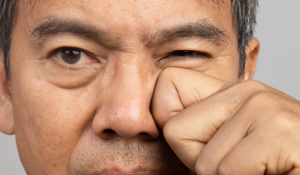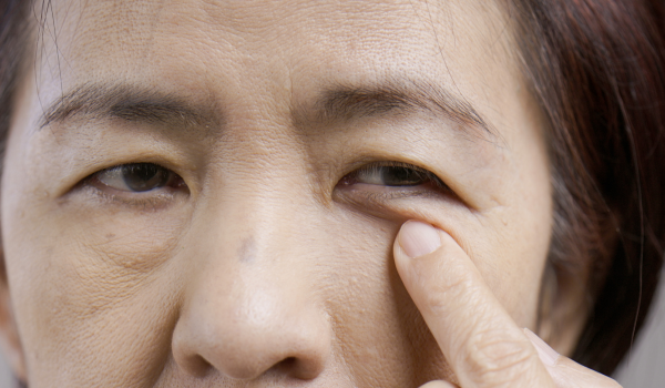
Ptosis, also known as blepharoptosis, is a medical condition characterized by a drooping upper eyelid. It occurs when the levator muscle, which is responsible for lifting the eyelid, becomes weakened or impaired. The severity of ptosis can vary widely. In mild cases, it may only affect appearance, but in more serious instances, the drooping can obstruct vision and interfere with daily life.
Though relatively rare, ptosis affects a small percentage of the population. It can be present at birth, known as congenital ptosis, or develop later in life, referred to as acquired ptosis. Congenital ptosis is more commonly seen in individuals assigned male at birth. Treatment depends on the type and severity of the condition and may involve surgery to correct the eyelid position and restore proper function.
Types of Ptosis
Ptosis is generally classified based on its cause. While congenital ptosis is the most common, acquired forms can result from nerve damage, muscle disorders, trauma, or aging.
congenital ptosis
This type is present at birth or develops during the first year of life. It accounts for the majority of ptosis cases in children.
neurogenic ptosis
Caused by nerve damage that affects the levator muscle. It may occur in conjunction with neurological disorders such as multiple sclerosis, third nerve palsy, or Horner syndrome.
myogenic ptosis
Results from muscle-related disorders, including myasthenia gravis or other conditions that affect the eye muscles and their function.
mechanical ptosis
Occurs when a physical mass such as a tumor, growth, or even prolonged contact lens use weighs down the eyelid and disrupts muscle function.
aponeurotic ptosis
Typically linked to aging, trauma, or eye surgery, this form occurs when the levator muscle becomes stretched or detached.
traumatic ptosis
Develops after direct injury to the eye or surrounding muscles, such as a blow or cut.
Symptoms
Ptosis symptoms depend on the severity of the eyelid drooping. While mild cases may not interfere with vision, moderate to severe ptosis can cause functional and cosmetic concerns.
common symptoms include:
-
Drooping of one or both upper eyelids
-
Uneven eyelid creases
-
Obstructed or reduced vision
-
Head tilting backward to see more clearly
-
Forehead wrinkles from raising eyebrows to compensate
-
Persistent eye fatigue or heaviness
-
Crossed eyes or lazy eye (amblyopia)
-
Difficulty focusing or double vision
-
Slight eyebrow lift while chewing
In most unilateral cases (only one eye affected), the left eyelid is more commonly involved.
What Causes Ptosis?
The eyelids are controlled by two key muscles: the levator and Müller’s muscle. When these muscles, especially the levator, become weak or underdeveloped, the upper eyelid begins to droop.
congenital causes
Congenital ptosis may result from improper development of the levator muscle or issues affecting the nerve that supplies it. This condition may be idiopathic (with no clear cause) or associated with other nerve-related disorders in infants, such as:
-
Marcus Gunn jaw-winking syndrome
-
Third cranial nerve palsy
-
Blepharophimosis syndrome
-
Horner’s syndrome
acquired causes
Acquired ptosis develops later in life and can be triggered by various factors, including:
-
Natural aging
-
Neurological conditions like multiple sclerosis or myasthenia gravis
-
Eye tumors or cysts
-
Repeated contact lens use
-
Eye trauma or injury
-
Post-surgical complications
-
Stroke or related nerve damage
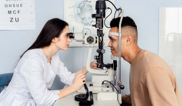
Diagnosis
Diagnosing ptosis involves evaluating the extent of eyelid drooping, identifying the cause, and ruling out similar conditions. After reviewing your medical history, your healthcare provider may perform several tests:
levator function test
Measures the eyelid’s upward movement while restricting the brow, to assess muscle strength.
margin-reflex distance (MRD)
Uses light reflection to calculate the space between the center of the pupil and the upper eyelid margin. A measurement below 4–5 mm typically indicates ptosis.
palpebral fissure height
Determines the vertical distance between upper and lower eyelids to assess symmetry and severity.
margin crease distance
Evaluates the distance between the eyelid margin and the crease above it while looking down. A higher than normal measure may indicate levator weakening.
additional tests
These may include thyroid function tests, vision tests, blood tests for autoimmune markers (such as acetylcholine receptor antibodies), and imaging (CT or MRI) if a mass is suspected.
Grading Ptosis
To plan treatment, ptosis is graded by its severity based on MRD (margin-reflex distance):
-
Mild: MRD difference of 2 mm or less
-
Moderate: MRD difference of 3 mm
-
Severe: MRD difference of 4 mm or more
These measurements help healthcare providers determine the best surgical or therapeutic approach.
Treatments for Ptosis
Treatment is generally required for moderate to severe cases that affect vision or appearance. The main objectives are to restore eyelid position, improve symmetry, and relieve obstruction.
Surgical treatments
levator resection
The levator muscle is shortened to lift the eyelid more effectively.
Matais procedure
The levator is connected to another eye muscle (superior rectus) to improve eyelid elevation.
Hess’s procedure
Involves attaching the levator to the frontalis muscle of the forehead.
frontalis suspension
In severe cases, a sling made of tissue or synthetic material connects the eyelid to the forehead muscle.
aponeurotic repair
Strengthens the levator's connection using synthetic reinforcements.
blepharoplasty
Removes excess eyelid skin to reduce sagging, often done alongside other ptosis surgeries.
Nonsurgical options
In specific cases, eyelid crutches can be attached to glasses to lift the eyelid. Eyelid taping is another temporary measure for managing obstruction.
Medications
A prescription eye drop called Upneeq (oxymetazoline) has been approved for adults with acquired ptosis. It works by stimulating Müller’s muscle, causing a temporary eyelid lift. This treatment is best suited for mild to moderate cases.
Prevention
There is no reliable method to prevent congenital ptosis. However, some exercises may help strengthen eyelid muscles, such as gently resisting eyelid closure while raising the eyebrow. Some clinicians recommend using the back of an electric toothbrush to stimulate the muscle, though evidence is limited.
Related Conditions
Ptosis during infancy or childhood may increase the risk of developing related complications:
-
Astigmatism: Pressure from drooping eyelids may distort the eye’s shape and affect vision.
-
Lazy eye (amblyopia): If left untreated, ptosis can weaken one eye’s vision permanently.
-
Torticollis: Infants may tilt or twist their head to see better, leading to abnormal neck posture.
Living With Ptosis
Ptosis can be managed successfully through timely treatment. While complications such as swelling or infection may occur after surgery, the procedure is generally effective. Most patients report high satisfaction rates following surgical correction.
In addition to the physical effects, ptosis may impact mental health due to changes in appearance. Anxiety, depression, or self-consciousness are not uncommon. Emotional support and counseling may be beneficial for those coping with these challenges.
Frequently Asked Questions
Does ptosis go away on its own?
Ptosis may remain constant, worsen, or fluctuate depending on the type and underlying cause. In some cases, treating the root condition may improve symptoms, but most moderate or severe cases require medical intervention.
What happens if ptosis is left untreated?
Mild ptosis may not require treatment, but untreated moderate or severe ptosis—especially in children—can lead to vision problems, astigmatism, lazy eye, or abnormal head posture.
Is ptosis surgery painful?
The procedure is usually performed under local anesthesia and is not painful. Some swelling or bruising may occur after surgery, but discomfort is typically manageable with medication.
How long is recovery after ptosis surgery?
Initial recovery takes a few days, with full healing usually occurring within two to three weeks. Patients are often asked to wear an eye patch for 24 hours and apply ice to reduce swelling.