.png)
Detecting skin cancer early can save lives. While some warning signs are visible to the naked eye, confirming whether a mole or patch is cancerous requires medical testing. Modern diagnostic tools allow dermatologists to distinguish harmless growths from dangerous ones—and determine the best treatment path.
This guide breaks down the main diagnostic steps, testing methods, and what to expect during the process, from your first skin check to biopsy results.
Why Diagnosis Matters
Early detection improves survival
When identified in its earliest stages, most skin cancers—including melanoma—can be treated effectively with minimal procedures. Delayed diagnosis, however, allows the cancer to grow deeper into the skin or spread to lymph nodes, making treatment more complex.
Visual inspection isn’t enough
While some moles or patches look obviously abnormal, many skin cancers mimic harmless spots. That’s why dermatologists rely on a combination of visual assessment, imaging tools, and laboratory tests to confirm what’s happening beneath the surface.
Initial Skin Examination
Comprehensive visual check
A skin cancer diagnosis usually begins with a full-body skin exam. Your dermatologist will inspect your entire skin surface, from scalp to toes, using bright lighting and magnification. They’ll look for:
-
New or changing moles
-
Non-healing sores
-
Scaly or crusted patches
-
Discolored or irregular growths
Reviewing personal and family history
You’ll also be asked about:
-
Past sunburns or tanning bed use
-
Family history of melanoma or skin cancer
-
Immune system disorders or prior radiation exposure
-
Medications that increase sun sensitivity
Photographic documentation
Dermatologists may take clinical photos for future comparison, especially if you have many moles or high-risk factors.
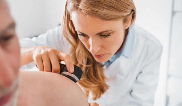
Dermatoscopy and Skin Imaging
What is dermatoscopy?
A dermatoscope is a handheld magnifying device that illuminates and enlarges the skin up to 10–20 times. It allows your doctor to see structures beneath the skin’s surface—such as pigment patterns, blood vessels, and texture—that aren’t visible to the naked eye.
Why it’s useful
Dermatoscopy helps distinguish between:
-
Benign moles and melanoma
-
Seborrheic keratosis vs. squamous cell carcinoma
-
Pigmented vs. non-pigmented lesions
It’s a non-invasive, painless procedure and plays a major role in improving diagnostic accuracy.
Digital mole mapping
For people with numerous moles, digital mole mapping combines high-resolution images with software that tracks changes over time. This technology helps identify subtle growth or color variations between visits—an early clue for melanoma detection.
When a Biopsy Is Needed
If a spot appears suspicious, your dermatologist will perform a skin biopsy—the gold standard for confirming skin cancer. This simple, usually outpatient procedure involves removing part or all of the lesion for microscopic examination.
Why biopsy is essential
No matter how experienced a doctor is, visual inspection alone can’t confirm cancer. The biopsy allows a pathologist to examine skin cells under a microscope, identifying whether they’re benign, precancerous, or malignant—and what type of cancer is present.
Types of Skin Biopsies
Different biopsy methods are chosen based on the lesion’s size, depth, and location.
Shave biopsy
-
Removes the top layers of skin using a scalpel or razor blade
-
Often used for superficial or raised lesions
-
Quick, minimal bleeding, no stitches required
Punch biopsy
-
Uses a circular tool to remove a small core of skin, including deeper layers
-
Ideal for diagnosing rashes or pigmented moles
-
Usually closed with one or two stitches
Excisional biopsy
-
Removes the entire lesion and a small margin of normal tissue
-
Commonly used when melanoma is suspected
-
Offers the most complete diagnostic sample
Incisional biopsy
-
Takes a portion of a large or difficult-to-remove lesion for testing
-
Used when removing the whole area isn’t feasible (e.g., on face or hand)
Your doctor will numb the area with local anesthesia, so you’ll feel little to no discomfort.
Laboratory Analysis
Once the tissue is removed, it’s sent to a pathology laboratory for microscopic examination by a dermatopathologist—a specialist trained in diagnosing skin diseases.
Microscopic examination
Under the microscope, the pathologist looks for:
-
Abnormal cell shapes or arrangements
-
Cancerous invasion into deeper layers
-
The type of skin cancer (basal cell, squamous cell, or melanoma)
-
Margins—whether cancer extends to the edge of the sample
Histologic staining
Special dyes, such as H&E (hematoxylin and eosin), highlight cellular details. Additional stains or molecular markers may be used to confirm ambiguous cases.
Report findings
Results typically arrive within 3–10 days. Your dermatologist will explain:
-
Whether the lesion is cancerous
-
What type and stage it is
-
Whether further testing or surgery is needed
Additional Diagnostic Tests
If the biopsy confirms skin cancer—especially melanoma—your doctor may order further tests to determine how far it has spread.
Sentinel lymph node biopsy
For melanomas thicker than 1 millimeter or with high-risk features, a sentinel lymph node biopsy checks whether cancer cells have reached nearby lymph nodes. A radioactive tracer or dye is injected near the tumor, guiding the surgeon to the first node that drains the area. If it’s cancer-free, the others are usually clear.
Imaging tests
Advanced imaging may include:
-
CT scan: checks internal organs or lymph nodes for spread
-
MRI scan: evaluates soft tissue or brain involvement
-
PET scan: detects metabolic activity of cancer cells throughout the body
-
Ultrasound: non-invasive method for regional lymph node evaluation
Blood tests
Although no blood test directly diagnoses skin cancer, bloodwork may help assess overall health before surgery or monitor treatment progress.
Understanding Staging
Once cancer is confirmed, doctors classify it into stages that describe how advanced it is. Staging guides treatment decisions and predicts prognosis.
Stage 0 (in situ)
Cancer cells are confined to the outermost skin layer (epidermis). Treatment is usually simple excision.
Stage I and II (localized)
Cancer has grown deeper into the skin but hasn’t spread to lymph nodes. Surgery is typically curative.
Stage III (regional spread)
Cancer has reached nearby lymph nodes or tissues. Additional surgery, radiation, or immunotherapy may be needed.
Stage IV (distant spread)
Cancer has metastasized to distant organs such as lungs, liver, or brain. Treatment focuses on systemic therapies like targeted drugs or immunotherapy.
Molecular and Genetic Testing
Personalized medicine in skin cancer
Advanced laboratories now analyze genetic mutations in tumor cells to guide targeted therapy.
Common molecular markers include:
-
BRAF and MEK mutations in melanoma (treated with targeted inhibitors)
-
PD-L1 expression for immunotherapy suitability
-
TP53 or PTCH1 mutations in basal and squamous cell carcinoma
These tests help determine which modern treatments will be most effective for your specific tumor type.
Second Opinions and Expert Review
Why second opinions matter
Pathology is precise, but interpretation can vary between specialists. A second opinion from another dermatopathologist ensures accuracy—especially for ambiguous or borderline results.
When to request one
-
If your diagnosis is rare or complex
-
If the treatment involves major surgery or systemic therapy
-
When results are unclear or inconsistent with clinical findings
Most labs can send slides and tissue blocks to other institutions upon request.
Emotional Impact of Diagnosis
It’s normal to feel anxious
Hearing the word “cancer” can trigger fear and confusion. Remember that skin cancer—especially when caught early—has some of the highest survival rates among all cancers.
Coping strategies
-
Ask detailed questions about your diagnosis and treatment
-
Bring a friend or family member to appointments
-
Join skin cancer support groups or counseling services
-
Focus on positive actions: treatment, prevention, and follow-up care
Knowledge is power—and peace of mind often begins with understanding your options.
Follow-Up After Diagnosis
Post-biopsy care
-
Keep the wound clean and covered for 24–48 hours
-
Avoid swimming until stitches are removed or the site heals
-
Follow your doctor’s instructions for scar management
Ongoing surveillance
After treatment, you’ll need regular skin checks:
-
Every 3–6 months for the first 2 years
-
Annually after that for life
Your dermatologist will monitor for recurrence and new lesions, especially if you’ve had melanoma or multiple skin cancers before.
Prevention After Diagnosis
Sun protection becomes essential
After a skin cancer diagnosis, protecting your skin becomes even more critical. Your remaining skin cells may already carry UV-induced mutations.
Smart protection habits
-
Apply SPF 30+ broad-spectrum sunscreen daily
-
Wear UPF clothing, hats, and sunglasses
-
Avoid tanning and peak sun hours
-
Perform monthly self-exams
Early detection prevents recurrence—and helps safeguard your future health.
Key Takeaways
-
Skin cancer diagnosis begins with visual inspection, often followed by biopsy.
-
Dermatoscopy and digital imaging improve early detection accuracy.
-
Biopsy and pathology confirm cancer type and stage.
-
Imaging and genetic tests assess spread and guide treatment.
-
Early detection ensures the best outcomes—never ignore a suspicious mole or patch.
Your skin tells your health story—listen to it carefully, and act early when something changes.


.png)
.png)
.png)
.png)
.png)
.png)
.png)
.png)
.png)