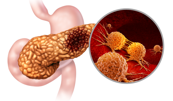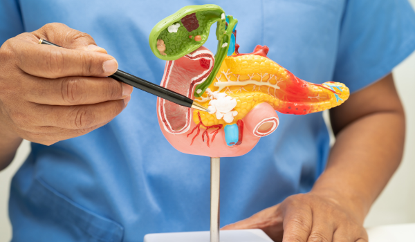
Initial Medical Assessment
Diagnosing pancreatitis begins with a detailed discussion between the patient and healthcare provider. The doctor will ask about the onset, duration, and intensity of symptoms, such as abdominal pain, nausea, and vomiting.
A comprehensive medical history helps identify possible triggers like gallstones, alcohol use, recent abdominal surgery, or medications. The provider will also ask about any previous episodes of similar symptoms, family history of pancreatic disease, and dietary patterns.
Physical examination focuses on abdominal tenderness, bloating, fever, or signs of jaundice. These early observations guide the selection of specific tests.
Physical Examination
During the physical exam, the healthcare provider will gently palpate the abdomen to identify tenderness, muscle guarding, or swelling.
Findings that may suggest pancreatitis include:
-
Upper abdominal pain that worsens when lying flat
-
Abdominal distension or firmness
-
Tenderness radiating toward the back
-
Fever and rapid heart rate
The examination may also include checking for skin changes, such as yellowing of the skin (jaundice) or bluish discoloration around the belly button (Cullen’s sign) and flanks (Grey Turner’s sign), which can indicate severe disease.
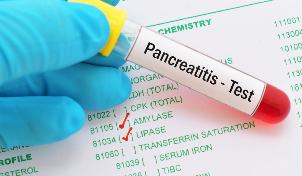
Blood Tests
Laboratory testing plays a crucial role in confirming pancreatitis. Key blood tests include:
Serum Amylase and Lipase:
-
These pancreatic enzymes are released into the bloodstream when the pancreas is inflamed.
-
Elevated lipase is considered more specific for pancreatitis, while amylase levels can rise in other abdominal conditions.
Complete Blood Count (CBC):
-
Detects elevated white blood cells, a sign of inflammation or infection.
Liver Function Tests (LFTs):
-
Helps determine if gallstones or bile duct obstruction are causing the pancreatitis.
Blood Glucose:
-
Measures how well the pancreas is regulating blood sugar.
-
High glucose levels can indicate impaired insulin production.
Electrolytes and Kidney Function Tests:
-
Assess dehydration and organ involvement.
Urine Tests
Urine tests may be ordered to evaluate kidney function and detect elevated amylase levels in cases where blood results are inconclusive.
Persistent high levels of urinary amylase can indicate ongoing pancreatic inflammation. Urine testing also helps rule out other abdominal or metabolic conditions.
Stool Tests
In chronic pancreatitis, stool analysis is important for evaluating fat malabsorption.
Tests such as fecal elastase measurement can confirm enzyme deficiency. Steatorrhea (pale, greasy stools) suggests advanced disease where the pancreas can no longer produce sufficient digestive enzymes.
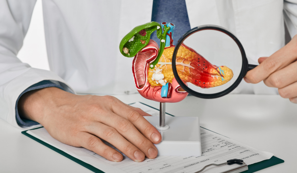
Abdominal Ultrasound
Ultrasound is often the first imaging test for suspected pancreatitis. It uses sound waves to create images of abdominal organs and is especially helpful for detecting gallstones, which are a leading cause of acute pancreatitis.
Benefits of ultrasound include:
-
No radiation exposure
-
Quick, non-invasive procedure
-
Can detect bile duct dilation or fluid around the pancreas
However, bowel gas and obesity can sometimes limit image clarity.
CT Scan
A computed tomography (CT) scan provides detailed cross-sectional images of the pancreas and surrounding tissues.
CT scans are essential for:
-
Confirming diagnosis when blood tests are unclear
-
Identifying complications like fluid collections, necrosis, or pseudocysts
-
Assessing the severity of inflammation
Contrast-enhanced CT is particularly valuable for evaluating blood flow and detecting tissue damage.
MRI and MRCP
Magnetic resonance imaging (MRI) and magnetic resonance cholangiopancreatography (MRCP) offer high-resolution imaging without radiation.
MRI can reveal subtle changes in pancreatic tissue, while MRCP focuses on bile and pancreatic ducts.
These tests are useful for detecting:
-
Ductal blockages
-
Pancreatic cysts or tumors
-
Chronic structural changes
Endoscopic Ultrasound (EUS)
EUS combines endoscopy with ultrasound for a closer look at the pancreas. A flexible tube with an ultrasound probe is inserted through the mouth into the stomach and duodenum, placing the probe near the pancreas for detailed images.
EUS is highly effective in:
-
Detecting small gallstones or sludge in the bile duct
-
Identifying early structural damage in chronic pancreatitis
-
Guiding fine-needle aspiration for biopsy
ERCP
Endoscopic retrograde cholangiopancreatography (ERCP) is both a diagnostic and therapeutic procedure. Using an endoscope, the doctor injects contrast dye into the bile and pancreatic ducts, followed by X-ray imaging.
ERCP can:
-
Identify blockages, strictures, or stones in the ducts
-
Remove gallstones or place stents to restore duct flow
While ERCP is highly effective, it carries risks, including the potential to cause pancreatitis itself, so it is usually reserved for cases where intervention is needed.
Genetic Testing
In patients with recurrent or chronic pancreatitis—especially those with a family history—genetic testing may identify mutations linked to inherited forms of the disease.
Mutations in genes like PRSS1, SPINK1, and CFTR can increase susceptibility, guiding prevention strategies and family counseling.
Biopsy
In rare cases, a biopsy of pancreatic tissue is performed to rule out cancer or other diseases that mimic pancreatitis.
Biopsy is typically guided by EUS or CT to ensure precise sampling with minimal risk.
Differential Diagnosis
Pancreatitis symptoms can resemble other abdominal conditions such as peptic ulcers, gallbladder disease, or bowel obstruction.
Physicians use test results in combination with symptom history to rule out other causes and confirm the diagnosis.
Severity Assessment
Once pancreatitis is confirmed, its severity must be evaluated to guide treatment.
Common scoring systems include:
-
Ranson’s Criteria (acute pancreatitis severity over 48 hours)
-
BISAP Score (predicts risk of complications)
-
APACHE II Score (used in critical care)
Severity assessment helps determine whether the patient can be managed in a general ward or requires intensive care.
Follow-Up Evaluations
After initial treatment, follow-up tests monitor recovery and check for complications. Repeat imaging may be needed if symptoms persist or worsen.
Long-term follow-up is especially important in chronic cases to prevent malnutrition and manage enzyme supplementation.
Final Thoughts
Diagnosing pancreatitis is a multi-step process that combines clinical evaluation, lab testing, and advanced imaging. Early and accurate diagnosis not only guides effective treatment but also helps prevent complications.
If you have symptoms such as persistent abdominal pain, nausea, or unexplained weight loss, seek prompt medical attention for a thorough evaluation.