.png)
Why Diagnosis Is Crucial
May-Thurner Syndrome (MTS), a condition where the left iliac vein is compressed by the right iliac artery, often goes undetected until it causes serious problems like deep vein thrombosis (DVT). Early detection through the right diagnostic tools is critical to prevent complications such as leg swelling, pain, skin discoloration, and life-threatening clots.
Because the symptoms of MTS mimic other conditions, diagnosis relies heavily on imaging and clinical evaluation. Understanding the step-by-step process helps patients and clinicians choose the best diagnostic strategy.
Initial Clinical Evaluation
Diagnosis usually starts with a visit to a primary care physician or vascular specialist. The doctor will assess your symptoms, medical history, and risk factors.
During the physical exam, they may check for:
-
Unilateral leg swelling (especially on the left)
-
Visible varicose veins or collateral veins
-
Discoloration or skin changes in the lower leg
-
Signs of DVT or previous clotting
Patients may be asked about family history, hormone use, prior surgeries, and pregnancy—all factors that can increase the likelihood of MTS.
Doppler Ultrasound
The first imaging step in evaluating suspected May-Thurner Syndrome is typically a Doppler ultrasound. This non-invasive test uses sound waves to visualize blood flow through veins and detect obstructions.
It’s most useful for identifying:
-
Presence of blood clots
-
Slow or turbulent blood flow in the deep veins
-
Vein diameter changes with position
However, Doppler ultrasound has limitations. It may miss iliac vein compression because these veins lie deep in the pelvis and are harder to assess from the body surface.
Duplex Ultrasound with Compression Techniques
Advanced ultrasound techniques may include duplex imaging, which combines real-time structure and flow visualization, and external compression methods to simulate vein response.
In skilled hands, this can raise suspicion of MTS if the blood flow significantly slows when pressure is applied or if abnormal vein diameter changes are noted. However, further imaging is often required for confirmation.
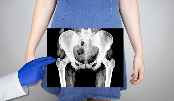
CT Venography
Computed Tomography (CT) venography offers a more detailed look at the pelvic vasculature. It uses contrast dye injected into a vein to highlight the iliac veins and arteries, allowing visualization of compressions or obstructions.
CT venography provides:
-
Cross-sectional imaging of the veins
-
Identification of vein narrowing and surrounding anatomy
-
Detection of clots or external masses compressing the vein
Its high resolution makes it useful in pre-operative planning and identifying the exact location and degree of compression.
MR Venography
Magnetic Resonance Venography (MRV) is a radiation-free alternative to CT. It uses magnetic fields and contrast agents to map the venous system in great detail.
MRV is particularly beneficial for:
-
Patients with contrast allergies or kidney issues (non-iodinated contrast is used)
-
Assessing blood flow dynamics over time
-
Identifying subtle vessel deformities or narrowing
However, MRV can be more expensive and less accessible, and it requires patients to remain still for extended periods during imaging.
Intravascular Ultrasound (IVUS)
IVUS is often considered the gold standard in diagnosing May-Thurner Syndrome. Unlike external ultrasound, this technique involves inserting a catheter with an ultrasound probe directly into the vein.
Benefits of IVUS include:
-
Direct visualization of the vein from the inside
-
Accurate measurement of vein diameter and stenosis
-
Ability to see fibrous bands or webs inside the vessel
This tool is typically used during venography or prior to interventions like stent placement.
Conventional Venography
Traditional venography involves injecting contrast dye into a vein and taking X-ray images as the dye flows through. This method allows doctors to visualize blockages or narrowing of the iliac veins.
Venography is useful for:
-
Confirming vein narrowing
-
Visualizing collateral veins that form around the blockage
-
Guiding catheter-directed procedures (e.g., thrombolysis)
While more invasive than other tests, venography remains an essential diagnostic tool, especially when combined with IVUS.
Cross-Sectional Imaging for Staging
To determine how far MTS has progressed, doctors may use cross-sectional imaging from CT or MR scans. This helps identify:
-
Stage 1: Asymptomatic compression
-
Stage 2: Symptomatic without thrombosis
-
Stage 3: Thrombosis or post-thrombotic syndrome
Staging guides treatment decisions and helps assess the urgency of intervention.
Differentiating from Other Conditions
One challenge in diagnosing MTS is that its symptoms overlap with other vascular or orthopedic problems. A thorough differential diagnosis is necessary to rule out:
-
Lymphedema
-
Peripheral artery disease
-
Sciatica or nerve impingement
-
Chronic venous insufficiency
Imaging is essential to confirm that symptoms are due to venous compression rather than unrelated disorders.
Laboratory Tests
Although imaging plays the primary role, lab tests may be ordered to support the diagnosis:
-
D-dimer: Elevated levels may suggest the presence of a blood clot
-
Coagulation profile: Assesses risk of clotting
-
Genetic testing: To detect inherited thrombophilias like Factor V Leiden
These tests don’t confirm MTS but help identify patients at higher risk of complications.
Role of Clinical Suspicion
MTS remains underdiagnosed in part because it requires a high level of clinical suspicion. Doctors should consider it if:
-
The patient has left-sided DVT without obvious cause
-
Leg symptoms worsen with prolonged sitting or standing
-
There are no visible clots, but imaging suggests reduced blood flow
-
Recurrent DVTs occur despite treatment
Awareness among clinicians significantly increases the likelihood of early and accurate diagnosis.
When to Refer to a Specialist
Patients suspected of having MTS should be referred to a vascular specialist or interventional radiologist if:
-
Standard imaging is inconclusive
-
Symptoms are persistent or worsening
-
Anticoagulants fail to improve the condition
-
There's a history of recurrent or unexplained DVT
Specialists can perform advanced imaging and offer minimally invasive treatment options.
Diagnosis in Special Populations
Different diagnostic approaches may be needed for certain groups:
-
Pregnant women: May require MRI instead of CT to avoid radiation
-
Young adults: Early-stage MTS may present with mild symptoms; IVUS can confirm subtle narrowing
-
Elderly patients: Diagnosis must rule out other age-related causes like heart failure or diabetes-related vascular disease
Tailoring the diagnostic process to individual risk factors and anatomy is key to accurate results.
Pediatric Diagnosis Challenges
Though rare in children, MTS can occur. Pediatric cases often involve congenital vein abnormalities or trauma. Diagnosis can be challenging because:
-
Symptoms may mimic growing pains
-
Imaging must be carefully adapted to the child’s size
-
Sedation may be required for advanced scans
Early detection in pediatric cases ensures better outcomes and minimizes long-term vascular damage.
Cost and Access Considerations
Not all diagnostic methods are available in every healthcare setting. Patients should:
-
Ask about insurance coverage for CT or MR venography
-
Discuss alternative methods if IVUS is unavailable
-
Seek second opinions if initial imaging is inconclusive
Timely and appropriate diagnosis often depends on both medical and logistical accessibility.
Avoiding Misdiagnosis
Misdiagnosis delays treatment and increases the risk of complications. To minimize errors:
-
Use a stepwise approach: begin with ultrasound, escalate to CT/MR/IVUS as needed
-
Don’t ignore symptoms that return after DVT treatment
-
Reassess if conservative treatment fails
-
Always consider anatomical factors when DVT appears without a clear cause
Clinicians should remain vigilant even when initial imaging doesn’t provide clear results.
Conclusion
Diagnosing May-Thurner Syndrome involves a combination of clinical awareness and advanced imaging techniques. From the initial Doppler ultrasound to sophisticated tools like IVUS and venography, each method contributes to a clearer picture of how blood flows through the pelvic veins—and where it gets obstructed.
Early and accurate diagnosis leads to better outcomes, prevents complications like DVT, and opens the door for minimally invasive treatment. Whether you’re a patient or healthcare provider, understanding the diagnostic pathway is the first step toward effective care.

.png)
.png)
.png)
.png)
.png)
.png)
.png)
.png)