Diagnosing psoriatic arthritis (PsA) can be challenging because there is no single test that can confirm the condition. Instead, healthcare providers rely on a combination of physical exams, blood tests, imaging studies, and medical history to reach an accurate diagnosis. The care team often includes a rheumatologist, who specializes in joints and bones, and a dermatologist, who focuses on skin health.
PsA is an autoimmune condition where the immune system mistakenly attacks healthy cells. This causes symptoms such as joint pain, skin inflammation, and swelling. Because PsA can progress rapidly, early testing is essential for effective management.
Medical History
understanding your background
Your healthcare provider may begin with a written or oral medical history questionnaire to understand your health background and family history. Common questions include:
-
Do you or any family members have psoriasis?
-
Does anyone in your family have psoriatic arthritis?
-
Have you experienced any recent infections or illnesses?
-
How long have your symptoms been present?
-
What specific symptoms are you noticing?
Providers may also ask about particular symptoms:
fatigue: Feeling tired is common for people with PsA. About one in five individuals experience extreme fatigue that impacts daily activities.
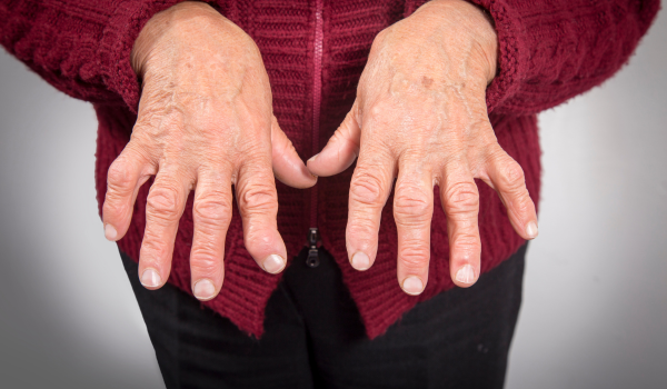
joint pain: Joint stiffness and soreness, especially in the morning, are hallmark signs. Swelling of the fingers and toes (dactylitis) or inflammation where tendons and ligaments attach to bones (enthesitis) are also frequent.
skin changes: Roughly one-third of those with psoriasis eventually develop PsA, often after several years of living with skin symptoms.
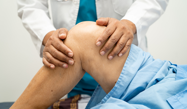
Physical Exam
examining visible and physical signs
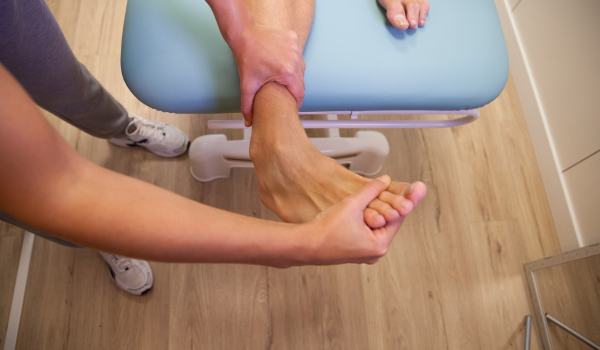
During the physical exam, a healthcare provider will assess joint appearance, flexibility, and any visible signs of inflammation. They may:
-
Look for skin discoloration, raised patches, or lesions
-
Check joints for stiffness, swelling, or tenderness
-
Compare symptoms on both sides of the body (as PsA often affects one side more than the other)
-
Measure vital signs such as blood pressure and heart rate
-
Press gently around affected areas to identify painful points
Since PsA can affect large joints and other areas, your provider may also examine your fingers, toes, nails, spine, and eyes for additional indicators of inflammation.
Imaging Tests
seeing the inflammation inside
If PsA is suspected, imaging tests help confirm joint inflammation and structural changes. Common options include:
computer tomography (CT) scan: Uses X-rays from multiple angles to create detailed images of bones and tissues, showing any joint damage or abnormal bone growth.
magnetic resonance imaging (MRI): Produces 3D images using magnets and radio waves. It can reveal soft tissue changes, inflammation, and early signs of bone damage.
ultrasound: A convenient and accessible test done in the doctor’s office. It detects soft tissue inflammation, fluid buildup, and bone erosion.
X-ray: Captures clear images of joints, useful in identifying long-term bone damage, though early PsA changes may not always appear on X-rays.
Blood Tests
checking for inflammation and antibodies
Blood tests help confirm PsA and rule out other similar conditions, such as rheumatoid arthritis. Several key tests may be ordered using a single blood sample:
anti-cyclic citrullinated peptide (Anti-CCP): Detects specific antibodies that attack healthy joint cells. Elevated levels may suggest advanced PsA.
C-reactive protein (CRP): Produced by the liver when inflammation occurs. Levels above 1.0 mg/dL may signal infection, autoimmune activity, or chronic inflammation.
erythrocyte sedimentation rate (ESR): Measures how quickly red blood cells settle in a blood sample. Faster rates often indicate higher inflammation levels.
human leukocyte antigen B27 (HLA-B27): A genetic marker linked to PsA and other autoimmune diseases. Abnormal results can point toward immune system dysfunction.
rheumatoid factor (RF): Helps differentiate PsA from rheumatoid arthritis. A negative result supports a PsA diagnosis since high RF levels are more typical of RA.
Routine follow-up blood tests may also be ordered to track inflammation and monitor treatment response.
Diagnostic Criteria
how doctors confirm PsA
Before 2006, PsA was often difficult to diagnose due to the lack of standardized criteria. That changed when an international group of rheumatologists developed the CASPAR (Classification Criteria for Psoriatic Arthritis) system.
The CASPAR criteria remain the most widely accepted tool for diagnosis. They assign point values to specific signs, symptoms, and test results. A score of three or more points indicates psoriatic arthritis.
| Category | Description | Points |
|---|---|---|
| Evidence of psoriasis | Current psoriasis (2 points) or personal/family history (1 point) | 1–2 |
| Psoriatic nail dystrophy | Nail pitting, thickening, or separation from the nail bed | 1 |
| Negative rheumatoid factor | Negative RF blood test | 1 |
| Dactylitis | Current or past swelling of an entire finger or toe | 1 |
| Radiographic evidence | Imaging showing new bone growth near joints | 1 |
These criteria are used both in clinical trials and everyday medical practice to help confirm a PsA diagnosis.


.png)