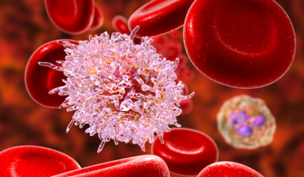
Leukemia is a cancer of the blood-forming tissues, often originating in the bone marrow. It impacts the development of red blood cells, white blood cells, and platelets. Common symptoms include fever, fatigue, unexplained bleeding, and easy bruising.
If leukemia is suspected, your healthcare provider will refer you to an oncologist— a doctor who specializes in diagnosing and treating cancer. The diagnostic process typically includes a combination of blood work, imaging, bone marrow sampling, and sometimes spinal fluid analysis.
Medical History and Physical Exam
initial evaluation: The diagnostic journey often begins with a review of your symptoms and overall health history. Your provider will ask detailed questions about fatigue, bleeding, infections, and other changes you've noticed.
physical examination: During the exam, they’ll inspect your skin, mouth, and eyes, and feel for swelling in the lymph nodes, liver, and spleen. Any signs of bruising, inflammation, or bleeding will also be assessed.
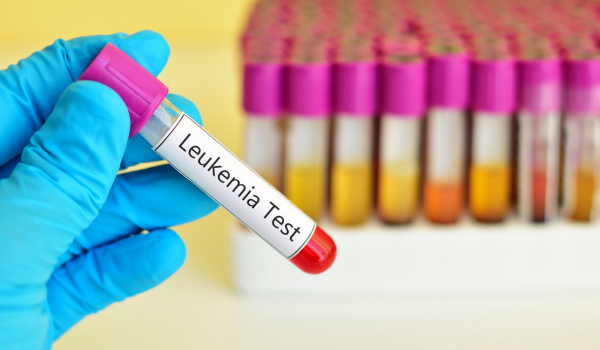
Blood Tests
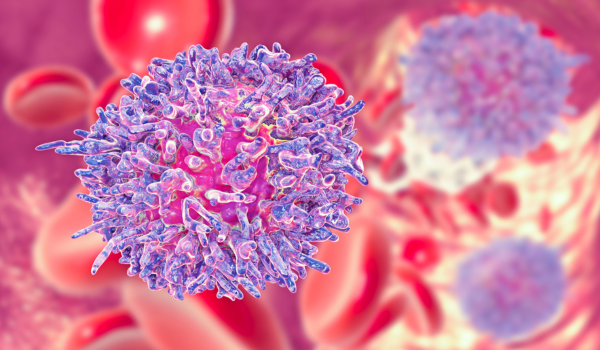
Leukemia alters the number and function of blood cells, which originate in the bone marrow. When cancer cells take over the marrow, healthy blood cell production declines.
affected blood cells include:
-
red blood cells (RBCs): carry oxygen from the lungs to tissues
-
white blood cells (WBCs): help the immune system fight infections
-
platelets: enable blood clotting to prevent excessive bleeding
tests used for diagnosis:
-
peripheral smear (blood smear): A lab examines your blood under a microscope to check for abnormal cell appearance or quantity.
-
complete blood count (CBC): Measures the levels of RBCs, WBCs, and platelets. High levels of immature white cells, like myeloblasts, may suggest leukemia.
-
comprehensive metabolic panel (CMP): Evaluates blood sugar, electrolytes, and organ function.
-
liver function tests (LFTs): Check for liver-related abnormalities.
-
coagulation panel: Assesses how well your blood clots, including fibrinogen levels.
Imaging Tests
Though leukemia doesn’t typically cause tumors, imaging may be used to assess for complications or infections caused by weakened immunity.
imaging options include:
-
x-ray: A chest x-ray may help identify lung infections.
-
computed tomography (CT) scan: Useful for evaluating lymph node enlargement and guiding biopsies.
-
magnetic resonance imaging (MRI): Detects leukemia involvement in the brain and spinal cord.
-
positron emission tomography (PET) scan: Tracks cancer spread by identifying sugar-absorbing cancer cells.
-
ultrasound: Non-invasive method to check for swollen organs or lymph nodes.
Bone Marrow Tests
Since leukemia begins in the bone marrow, direct sampling is essential for diagnosis.
common procedures:
-
bone marrow aspiration: A thin needle extracts liquid marrow, usually from the pelvic bone.
-
bone marrow biopsy: A slightly larger needle collects a small core of bone and marrow.
These procedures are done under local anesthesia and may cause brief discomfort.
Cytochemistry
Cytochemistry uses stains and dyes to detect changes in cells. Specific leukemia cells respond to these chemicals, aiding in identifying cancer types.
techniques include:
-
flow cytometry and immunohistochemistry: Identify leukemia cells by exposing them to antibodies and analyzing their response under a microscope.
-
chromosome and gene tests: Examine DNA and chromosomes for mutations often linked to specific types of leukemia.
Spinal Fluid Tests
To check if leukemia has spread to the central nervous system, a lumbar puncture may be recommended.
lumbar puncture (spinal tap): A small sample of cerebrospinal fluid is collected using a needle inserted into the lower back. The fluid is then examined by a pathologist to check for cancer cells.
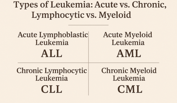
Staging of Leukemia
Unlike many other cancers, leukemia does not form tumors, so staging is based on blood and marrow cell counts rather than tumor size or spread.
staging for chronic lymphocytic leukemia (CLL): The Rai system is most commonly used in the U.S.
-
stage 0: High lymphocyte count; normal RBC and platelet levels; no swelling in lymph nodes, spleen, or liver
-
stage 1: High lymphocytes and enlarged lymph nodes
-
stage 2: High lymphocytes with enlarged liver or spleen
-
stage 3: High lymphocytes with anemia; platelets remain normal
-
stage 4: High lymphocytes with low platelet count; anemia may or may not be present
phases of chronic myeloid leukemia (CML):
-
chronic phase: Fewer than 10% blast cells; often no symptoms
-
accelerated phase: 10–19% blasts; symptoms like fever or weight loss may appear
-
blast phase: 20% or more blasts; more aggressive with worsening symptoms
other types of leukemia:
Staging for acute lymphocytic leukemia (ALL) and acute myeloid leukemia (AML) is based on blast cell counts and other lab results, though there is no universal staging system.
Screening for Similar Conditions
Several disorders share symptoms with leukemia. To ensure an accurate diagnosis, your provider may rule out other diseases such as:
-
viral infections: Viruses like HIV or Epstein-Barr virus can mimic leukemia by altering white blood cell levels
-
myeloproliferative disorders: Includes conditions like polycythemia vera that affect blood cell production
-
aplastic anemia: A condition where the marrow stops making blood cells
-
myelodysplastic syndromes (MDS): Pre-leukemia disorders that cause ineffective blood cell production
A Quick Review
Leukemia is a type of cancer affecting blood and bone marrow. Diagnosis involves a series of steps, including medical history, physical exams, blood work, imaging, bone marrow biopsies, and cytogenetic studies.
Staging is essential for treatment planning and varies by leukemia type. Since symptoms can overlap with other conditions, comprehensive testing helps confirm diagnosis and rule out alternatives.

.png)