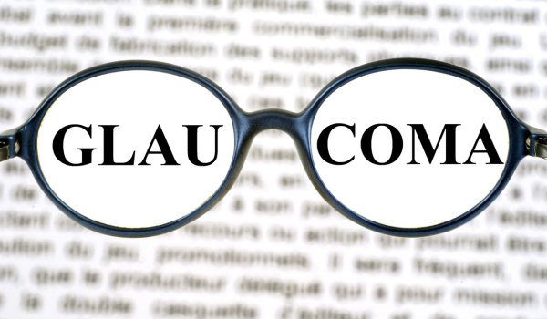
Glaucoma is a group of eye conditions that damage the optic nerve—the bundle of fibers responsible for transmitting visual information from the eye to the brain. The damage typically occurs when fluid builds up in the eye and raises intraocular pressure, eventually leading to irreversible vision loss or blindness.
Since glaucoma often develops without early symptoms, regular eye exams with a qualified specialist are essential for early detection and management.
Optometrists (eye care professionals who diagnose and treat eye disorders) and ophthalmologists (medical doctors trained to treat eye conditions and perform surgeries) are qualified to diagnose glaucoma.
To confirm a diagnosis, your eye specialist will perform a comprehensive eye examination that includes several important components:
Medical history review
Your provider will begin by reviewing your overall health and eye history to identify possible glaucoma risk factors. They may ask if you’ve experienced any of the following:
-
existing medical conditions such as high blood pressure, diabetes, or sleep apnea
-
past eye injuries
-
current symptoms like blurry vision, eye pain, or headaches
-
family history of glaucoma
-
long-term use of corticosteroids, including medications like prednisone or cortisone
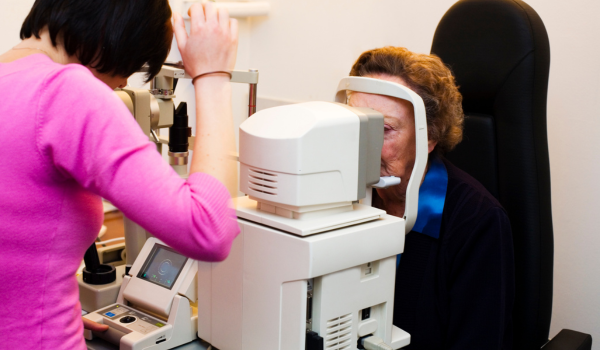
Eye pressure test (tonometry)
Tonometry measures intraocular pressure (IOP), a critical factor in glaucoma. Your eyes will be numbed with drops to reduce discomfort during the test.
There are several methods used to measure IOP:
-
goldmann applanation method: A small device gently flattens the cornea to determine eye pressure and is considered the gold standard for glaucoma monitoring.
-
electronic indentation tonometry: A handheld tool (tono-pen) touches the cornea to quickly measure pressure.
-
non-contact (air puff) method: A puff of air is used to flatten the cornea and estimate pressure without physical contact.
Each person’s normal eye pressure may vary, though typical readings range from 10 to 21 mmHg. While elevated pressure is a common sign of glaucoma, it’s possible to have glaucoma even within this range.
Cornea measurement (pachymetry)
Pachymetry measures the thickness of the cornea. A small probe gently touches your eye to perform the reading.
This measurement is important because corneal thickness can influence the accuracy of eye pressure readings. A thin cornea is also considered an independent risk factor for glaucoma.
Visual field test (perimetry)
A visual field test assesses your peripheral vision, which is often affected first in glaucoma. You’ll be asked to rest your chin on a bowl-shaped device while one eye is tested at a time.
As small lights appear in your side vision, you’ll press a button whenever you see them. This process helps map your full visual field and highlight any blind spots or narrowing of vision.
Results are compared to standard charts to assess the extent of vision loss and determine the severity of glaucoma.
Back-of-the-eye examination (ophthalmoscopy)
Ophthalmoscopy allows your provider to inspect the optic nerve directly. Eye drops will be used to dilate your pupils, making it easier to see the back of the eye.
Using a small handheld device called an ophthalmoscope, your provider will look for signs of optic nerve damage, including a condition called “cupping,” where the center of the optic nerve appears enlarged—a classic indication of glaucoma.
Optical coherence tomography (OCT)
OCT is an imaging test that uses light waves to produce detailed cross-sectional images of your retina and optic nerve. During the scan, you’ll rest your head on a support while the machine scans your eyes painlessly.
This test measures the thickness of the nerve fiber layer and helps detect damage before noticeable vision loss occurs.
Eye drainage angle examination (gonioscopy)
Gonioscopy evaluates the drainage angle—the area where fluid leaves the eye. After numbing your eye, your specialist will place a handheld lens with a mirror onto your eye to view the angle between your cornea and iris.
If the drainage angle is narrow or blocked, fluid can’t exit properly, increasing the risk of glaucoma. Gonioscopy helps determine whether glaucoma is open-angle or angle-closure type.
A quick review
Glaucoma is a progressive condition that causes damage to the optic nerve and can lead to blindness if untreated. Because early stages often lack noticeable symptoms, routine eye exams play a critical role in early diagnosis.
Even if you have no vision complaints, undergoing regular eye screenings that include pressure checks, visual field tests, and imaging studies can help detect glaucoma early—offering the best chance to preserve your vision long-term.