.png)
Introduction
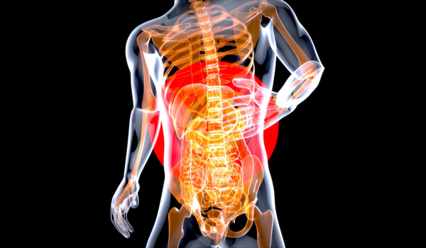
Stomach cancer, or gastric cancer, is often challenging to diagnose in its early stages due to vague or mild symptoms that can resemble common digestive issues. Early detection plays a critical role in improving survival rates and treatment outcomes. Once suspected, a series of diagnostic tests and procedures are used to confirm the presence of cancer, understand its characteristics, and determine the extent to which it has spread. This process is known as diagnosis and staging. Understanding how stomach cancer is diagnosed and staged can help patients and caregivers make informed decisions about care and treatment.
Initial Assessment and Medical History
The diagnostic journey typically begins with a visit to a healthcare provider. During this appointment, the doctor gathers a detailed medical history, including symptoms, lifestyle factors, dietary habits, personal and family history of cancer, and any past medical conditions such as ulcers or chronic gastritis. The physical examination may include checking the abdomen for tenderness, swelling, or palpable masses.
Although the initial assessment cannot confirm cancer, it provides important clues that may prompt further investigation. If stomach cancer is suspected, more specific diagnostic tools are required.
Endoscopy and Biopsy
The gold standard for diagnosing stomach cancer is an upper endoscopy, also known as esophagogastroduodenoscopy (EGD). In this procedure, a thin, flexible tube with a light and camera is inserted through the mouth and into the stomach. This allows doctors to directly visualize the lining of the stomach and detect any abnormal areas, such as ulcers, tumors, or inflammation.
If suspicious tissue is observed, a biopsy is performed during the endoscopy. This involves removing a small sample of tissue for examination under a microscope by a pathologist. A biopsy is the only definitive method to confirm the presence of cancer cells.
Imaging Tests
Once cancer is confirmed, imaging tests are used to determine the extent of the disease and whether it has spread to other areas of the body. Common imaging techniques include:
-
Computed Tomography (CT) Scan: A CT scan provides cross-sectional images of the abdomen and pelvis, helping to identify tumors, lymph node involvement, and metastases to organs like the liver or lungs.
-
Magnetic Resonance Imaging (MRI): MRI uses magnetic fields and radio waves to create detailed images, especially useful for evaluating soft tissues and blood vessels.
-
Positron Emission Tomography (PET) Scan: A PET scan detects cancerous cells based on their metabolic activity. It is often combined with CT (PET-CT) to provide both functional and anatomical information.
-
Upper Gastrointestinal (GI) Series: This involves swallowing a contrast material (usually barium) that coats the stomach lining, followed by X-rays. While less commonly used today, it can help detect abnormalities in the stomach’s shape or movement.
Endoscopic Ultrasound (EUS)
Endoscopic ultrasound is a specialized test that combines endoscopy with ultrasound technology. During this procedure, an ultrasound probe attached to an endoscope is inserted into the stomach. This allows for detailed imaging of the stomach wall and nearby lymph nodes.
EUS is particularly valuable for determining the depth of tumor invasion into the stomach wall and assessing whether nearby lymph nodes are involved. It is often used for more accurate staging of early gastric cancers and for guiding fine-needle aspiration biopsies of lymph nodes or other suspicious areas.
Laparoscopy
In some cases, laparoscopy is performed to gain a clearer view of the abdominal cavity. This minimally invasive surgical procedure involves inserting a small camera through a tiny incision in the abdomen. It allows doctors to detect small tumors or metastases that might not be visible on imaging scans.
Laparoscopy can also be used to collect fluid or tissue samples from the abdominal cavity for further testing. It is often recommended before major surgery to ensure that the cancer has not spread beyond what imaging has detected.
Laboratory Tests
Blood tests are not used to diagnose stomach cancer directly but can provide helpful information about a patient’s overall health. These may include:
-
Complete Blood Count (CBC): To check for anemia, which may result from internal bleeding or poor nutrition.
-
Liver Function Tests: To evaluate whether the liver has been affected by cancer.
-
Tumor Markers: Substances like CEA (carcinoembryonic antigen) and CA 19-9 can sometimes be elevated in stomach cancer but are not specific or reliable enough for diagnosis. However, they may be used to monitor response to treatment.
Staging the Cancer
Once stomach cancer is diagnosed, determining its stage is crucial for developing an effective treatment plan. Staging describes how far the cancer has spread and helps predict prognosis. The most commonly used system is the TNM staging system, which classifies cancer based on:
-
T (Tumor): How deeply the tumor has invaded the stomach wall.
-
N (Nodes): Whether nearby lymph nodes contain cancer cells.
-
M (Metastasis): Whether cancer has spread to distant organs.
The combination of these factors leads to an overall stage from Stage 0 (very early) to Stage IV (advanced):
-
Stage 0: Abnormal cells are present but confined to the innermost layer.
-
Stage I: Cancer has begun to invade deeper layers of the stomach wall.
-
Stage II: Cancer has spread to more lymph nodes or further into the stomach.
-
Stage III: Cancer has spread extensively within the stomach and nearby nodes.
-
Stage IV: Cancer has metastasized to distant organs.
Accurate staging is essential for choosing the appropriate treatment, which may include surgery, chemotherapy, radiation therapy, or a combination of these options.
The Importance of Multidisciplinary Evaluation
Stomach cancer diagnosis and staging often require a team approach. A multidisciplinary team may include gastroenterologists, oncologists, radiologists, pathologists, and surgeons. Each specialist contributes their expertise to ensure the diagnosis is accurate and that the most effective treatment strategy is chosen.
Regular team meetings or tumor boards help coordinate care, especially for complex or advanced cases. This approach also ensures that patients have access to the latest treatments and clinical trials.
Conclusion
Diagnosing and staging stomach cancer is a detailed process that involves various procedures and technologies. From endoscopy and biopsy to imaging and surgical exploration, each step plays a role in identifying the disease and understanding its extent. Accurate diagnosis and staging are the foundation of personalized treatment and improved outcomes.
As with many cancers, early detection remains key. Individuals experiencing persistent digestive symptoms should seek medical attention without delay. Increased awareness, advanced diagnostic tools, and a collaborative approach to care are vital components in the fight against stomach cancer.
.png)
.png)
.png)
.png)
.png)