.png)
Introduction
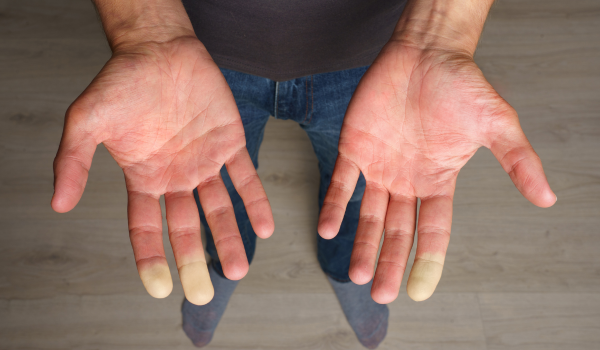
Raynaud’s Phenomenon involves sudden and temporary narrowing of blood vessels, leading to reduced blood flow to the extremities. This vascular reaction explains the characteristic color changes in the fingers and toes. Understanding the underlying vascular changes is crucial for recognizing the complexity of this condition and the physiological processes that drive its symptoms.
Normal Vascular Function
Under typical conditions, blood vessels respond dynamically to temperature changes and physical demands. The autonomic nervous system regulates vasoconstriction and vasodilation to maintain consistent internal temperature and organ perfusion. In Raynaud’s, this system becomes overly reactive, resulting in exaggerated responses to cold or stress.
Vasospasm and Reduced Blood Flow
The primary vascular abnormality in Raynaud’s is vasospasm, which causes blood vessels to constrict suddenly. These spasms typically affect the small arteries and arterioles in the fingers and toes. During a Raynaud’s episode, blood supply is temporarily cut off, leading to pallor (whiteness), followed by cyanosis (blue color) as oxygen levels drop. When the spasm subsides, a rush of blood returns, resulting in redness and throbbing pain.
Role of Endothelial Dysfunction
The endothelium is the inner lining of blood vessels and plays a critical role in vascular health. In people with Raynaud’s, this lining may be impaired, reducing the ability of blood vessels to dilate. Endothelial dysfunction may also contribute to inflammation and increased sensitivity to stress hormones.
Increased Sympathetic Nervous System Activity
The sympathetic nervous system controls the body’s fight-or-flight response. In Raynaud’s, this system becomes hyperactive, triggering vasospasm even with minor cold exposure or emotional stress. This response is particularly evident in primary Raynaud’s but may also contribute to secondary forms.
Structural Vascular Changes in Secondary Raynaud’s
Secondary Raynaud’s is associated with chronic conditions like scleroderma or lupus, which can cause structural damage to blood vessels. These changes may include thickening of vessel walls, fibrosis, and permanent narrowing. As a result, blood flow may be persistently reduced even between attacks.
Microvascular Abnormalities
Capillary abnormalities, such as dilated or irregular capillaries, have been observed in individuals with secondary Raynaud’s. These changes can be identified using nailfold capillaroscopy and may provide insight into disease severity and progression.
Other Contributing Mechanisms
-
Platelet Activation: Enhanced platelet aggregation can increase the risk of clotting and impair blood flow.
-
Oxidative Stress: The production of reactive oxygen species may damage vascular tissue, contributing to dysfunction.
-
Cold-Induced Protein Responses: Cold exposure may increase levels of proteins that affect blood flow regulation.
Conclusion
Raynaud’s Phenomenon is a condition driven by complex vascular mechanisms, primarily involving exaggerated vasospasm, endothelial dysfunction, and sympathetic nervous system overactivity. In secondary cases, structural damage to blood vessels adds another layer of complexity. By understanding these vascular changes, healthcare providers and patients can better target treatments and management strategies to improve quality of life.

.png)


.png)
.png)